We use cookies to improve your browsing experience and provide meaningful content. Read our cookie policy. Accept
  •  Customer Login
  • Register
  •  View Cart (0)
  •  Customer Login
  • Register
  •  View Cart (0)

  • Products
  • Services & Support
  • Learning centers
  • APPLICATIONS
  • About
  • Contact Us

Close

  • ‹ Back to ProteoTuner protein control systems
  • Plasmid systems
  • Viral systems
  • ProteoTuner antibody
  • Shield1 ligand
Overviews ProteoTuner explained
Learn more about our tet, ProteoTuner, and iDimerize inducible systems Inducible systems
Home › Products › Gene function › ProteoTuner protein control systems › Viral systems

Gene function

  • Gene editing
    • CRISPR-Cas9
      • Long ssDNA for knockins
      • Knockin screening kit
      • Genome-wide sgRNA library system
      • Recombinant Cas9 protein
      • GMP recombinant Cas9
      • Mutation detection kits
      • In vitro transcription and screening kits
      • Cas9-sgRNA gesicle production
      • Cas9 antibodies
      • Plasmid systems
      • Lentiviral systems
      • AAV systems
      • RNA transfection
      • Genotype confirmation kit
      • Indel identification kit
    • Cre recombinase
      • AAV2-Cre recombinase
      • Cre Recombinase Gesicles
      • Cre recombinase RLPs
  • Viral transduction
    • Adeno-associated virus (AAV)
      • Vector systems
        • Helper-free expression system (CMV promoter)
        • Tet-inducible promoter
        • CRISPR/Cas9 system
        • Cre recombinase system
        • Beta-galactosidase system
        • ZsGreen1 control vector
      • qPCR titration
      • Purification kits
      • AAV concentration
      • Packaging systems and cells
        • 293T cell line
        • Extraction solutions
        • Packaging plasmid sets
    • Adenovirus
      • Vectors and packaging
        • Expression system 3
        • Tet inducible
        • Adeno-X 293 cells
      • CAR Receptor Booster
      • Purification kits
      • Titration kits
    • Lentivirus
      • Vector systems
        • Constitutive promoter
        • EF-1 alpha promoter
        • IRES bicistronic
        • Fluorescent protein
        • Tet-inducible
      • Premade lentiviral particles
        • Whole-cell labeling
        • Organelle labeling
        • Tet-On 3G transactivator
      • Packaging systems and cells
        • Lenti-X packaging single shots
        • Lenti-X 293T cells
      • Titration kits
        • Lenti-X GoStix Plus
        • qRT-PCR
        • p24 ELISA
        • Integrated copy number
      • Lentivirus concentration
      • Purification kits
      • Integration site analysis
      • Transduction enhancers
        • Lenti-X Accelerator
        • Ecotropic Receptor Booster
    • Retrovirus
      • Vector systems
        • Constitutive promoter
        • Fluorescent protein
        • Tet inducible
        • MSCV system
      • Packaging systems and cells
      • Titration kits
      • Retro-X Concentrator
      • Integration site analysis
      • Receptor booster
  • Fluorescent proteins
    • Fluorescent protein plasmids
      • Cyan and green fluorescent proteins
        • AcGFP1 fluorescent protein
        • ZsGreen1 fluorescent protein
        • GFP & GFPuv fluorescent proteins
        • AmCyan1 fluorescent protein
      • Red fluorescent proteins
        • mCherry fluorescent protein
        • DsRed-Monomer fluorescent protein
        • DsRed2 fluorescent protein
        • DsRed-Express and DsRed-Express2 fluorescent proteins
        • tdTomato fluorescent protein
        • AsRed2 fluorescent protein
        • mStrawberry fluorescent protein
      • Far-red fluorescent proteins
        • E2-Crimson fluorescent protein
        • HcRed1 fluorescent protein
        • mRaspberry fluorescent protein
        • mPlum fluorescent protein
      • Orange and yellow fluorescent proteins
        • mOrange2 fluorescent protein
        • mBanana fluorescent protein
        • ZsYellow1 fluorescent protein
      • Photoactivatable and photoswitchable proteins
        • Dendra2 fluorescent protein
        • Timer fluorescent protein
        • PAmCherry fluorescent protein
    • Subcellular labeling plasmids
    • Flow cytometer calibration beads
    • Recombinant fluorescent proteins
  • T-cell transduction and culture
    • RetroNectin reagent
    • LymphoONE T-cell medium
    • Cytokine GoStix Plus assays
    • Anti-CD3 antibody (OKT3)
    • CultiLife culture bags
    • RetroNectin ELISA kit
  • Tet-inducible expression systems
    • Tet-One systems
    • Tet-On 3G systems
      • Tet-On 3G systems
      • Tet-On 3G—lentiviral
      • Tet-On 3G—retroviral
      • Adeno-X Tet-On 3G inducible expression system
      • Tet-On 3G cell lines
      • Tet-On 3G systems—bidirectional
      • Tet-On 3G systems—bicistronic
      • Tet-On 3G systems—EF1-alpha promoter
    • Tet systems legacy products
      • Tet-Off cell lines
      • Tet-On and Tet-Off—2nd generation
      • Tet-tTS transcriptional silencer
    • Tet-inducible miRNA systems
    • TetR monoclonal antibody
    • Tet-approved FBS
  • ProteoTuner protein control systems
    • Plasmid systems
    • Viral systems
    • ProteoTuner antibody
    • Shield1 ligand
  • iDimerize inducible protein interaction systems
    • Homodimerization systems
    • Heterodimerization systems
    • Reverse dimerization
    • Regulated transcription
    • Dimerizer ligands
    • In vivo applications
    • Dimerization domain antibodies
  • Transfection reagents
    • Plasmid transfection reagents
      • Xfect reagent
      • Xfect for mES cells
      • Calcium phosphate transfection
      • Fluorescent transfection controls
    • Protein transfection reagents
    • RNA transfection reagents
  • Mammalian expression plasmids
    • Plasmids with selectable markers
    • Bidirectional promoter vectors
    • Bicistronic IRES vectors
    • MicroRNA expression
  • Cell biology assays
    • Extracellular vesicle isolation
    • Reporter systems
      • Fluorescent protein promoter reporters
        • Promoterless (lowest background)
        • Promoterless (traditional)
      • Proteasome monitoring
      • Secreted alkaline phosphatase assays
      • Beta galactosidase and LacZ vectors
      • Secreted luciferase assay
      • Fucci cell-cycle vectors
      • Lenti-X Actin Dynamics Monitoring Kit
    • Apoptosis detection kits
      • Annexin V apoptosis assay
      • PARP antibody
      • ApoAlert caspase assays
      • Apoptosis analysis
      • In situ apoptosis detection
    • Epigenetics
      • DNA methylation
        • EpiXplore Methylated DNA Enrichment Kit
        • EpiScope MSP Kit
        • EpiScope Nucleosome Preparation Kit
        • EpiTaq HS
        • EpiScope control DNA
    • Cell biology reagents
      • WST-1 cell proliferation
      • Heme oxygenase-1
    • RNA interference
      • miRNA
        • MicroRNA quantitation
      • siRNA
        • siRNA quantitation
    • Cell-culture accessories
      • Magnetic separator for cell culture
      • Antibiotics for cell biology
        • Anhydrotetracycline
      • Antibiotic selection markers & plasmids
    • Signal transduction
      • Pathway profiling vectors
      • Dominant negative vectors
Need help?
Contact Sales
Overviews ProteoTuner explained
Learn more about our tet, ProteoTuner, and iDimerize inducible systems Inducible systems

ProteoTuner expression systems in lentiviral and retroviral formats

ProteoTuner inducible expression systems in lentiviral and retroviral formats

Lentiviral and retroviral ProteoTuner systems make it possible to investigate the function of a specific protein of interest directly—by rapidly changing the abundance of the protein itself. These systems utilize a ligand-dependent destabilization domain (DD), and the ligand Shield1 to reversibly stabilize and destabilize a DD-tagged protein of interest in a predictable and dose-dependent manner. Your protein of interest is fused to a DD tag and rapidly stabilized by adding the Shield1 ligand to the culture medium. Plasmid formats are also available.

Cat. # Product Size License Quantity Details
632175 Lenti-X™ ProteoTuner™ Shield System N (w/ ZsGreen1) Each USD $740.00

License Statement

ID Number  
55 cPPT Element. This product and its use are the subject to one or more of the following U.S. Pat. No. 8,093,042. The purchase of this product conveys to the buyer the non-transferable right to use the purchased amount of the product and components of the product in research conducted by the buyer (whether the buyer is an academic or for-profit entity). The buyer cannot disclose information, sell or otherwise transfer this product, its components or materials made using this product or its components to a third party or otherwise use this product or its components or materials made using this product or its components for any commercial purposes. If the buyer is not willing to accept the limitations of this limited use statement, Takara Bio USA, Inc. is willing to accept return of the product with a full refund. For information on purchasing a license to the DNA-Flap technology for purposes other than research, contact the Transfer of Technology Office, Institut Pasteur, 28 rue du Docteur Roux, 75 724 Paris Cedex 15 (www.pasteur.fr).
57 This product is covered by U.S. Patent Nos. 8,173,792 and 9,487,787.
K11 This product and its use are the subject to one or more of the following U.S. Pat. 8,093,042 and foreign equivalents. The purchase of this product conveys to the buyer the non-transferable right to use the purchased amount of the product and com­ponents of the product in research conducted by the buyer (whether the buyer is an academic or for-profit entity). The buyer cannot disclose information, sell or otherwise transfer this product, its components or materials made using this product or its components to a third party or otherwise use this product or its components or materials made using this product or its components for any com­mercial purposes. If the buyer is not willing to accept the limitations of this limited use statement, Takara Bio USA is willing to accept return of the product with a full refund. For information on purchasing a license to the DNA-Flap technology for purposes other than research, contact the Transfer of Technology Office, Institut Pasteur, 28 rue du Docteur Roux, 75 724 Paris Cedex 15 (www.pasteur.fr).

The Lenti-X ProteoTuner Shield System N (w/ ZsGreen1) provides a unique method for quickly and directly regulating the amount of your protein of interest present in a cell, combined with versatile lentiviral delivery. The system utilizes a ligand-dependent destabilization domain (DD), derived from a 12 kDa mutant of the FKBP protein, and a membrane permeable stabilizing ligand, Shield1. Once cloned into the pLVX-PTuner Green Vector, the protein of interest is expressed with an N-terminal DD-tag and the fluorescent protein ZsGreen1. In the presence of Shield1, the DD-tagged protein is stabilized and will accumulate inside the cell. This ligand-dependent stabilization occurs very quickly, and has been observed as soon as 15–30 minutes after the addition of Shield1. In the absence of Shield1, the DD-tagged protein of interest is unstable. Consequently, removal of Shield1 (by splitting the cells into medium without Shield1) allows for destabilization and rapid degradation of the protein of interest. The extent of stabilization via Shield1 directly correlates with the amount of the ligand in the medium, so it is possible to fine-tune the amount of protein of interest present in the cell by controlling the amount of the Shield1 ligand.

Notice to purchaser

Our products are to be used for Research Use Only. They may not be used for any other purpose, including, but not limited to, use in humans, therapeutic or diagnostic use, or commercial use of any kind. Our products may not be transferred to third parties, resold, modified for resale, or used to manufacture commercial products or to provide a service to third parties without our prior written approval.

Documents Components Image Data Resources

Back

Schematic of lentiviral ProteoTuner vectors

Schematic of lentiviral ProteoTuner vectors
Schematic of lentiviral ProteoTuner vectors.

Back

632175: Lenti-X ProteoTuner Shield System N (w/ ZsGreen1)

632175: Lenti-X ProteoTuner Shield System N (w/ ZsGreen1)

Required Products

Cat. # Product Size Price License Quantity Details
632189 Shield1 500 uL USD $360.00

The Shield1 compound stabilizes a protein of interest tagged with the destabilization domain (DD), which is based on a mutated version of the FKBP protein. It is used to protect the DD fusion protein of interest from proteasomal degradation. The amount of Shield1 can be varied to tune the amount of stabilized protein of interest within the cell.

Documents Components Image Data

Back

DD fusions exhibit predictable, dose-dependent protein stabilization by Shield1; using the DD optimal for your protein is crucial in getting the best performance

 DD fusions exhibit predictable, dose-dependent protein stabilization by Shield1; using the DD optimal for your protein is crucial in getting the best performance

DD fusions exhibit predictable, dose-dependent protein stabilization by Shield1; using the DD optimal for your protein is crucial in getting the best performance. While DD-N (solid blue line) is suitable for N-terminal fusions and even C-terminal fusions for many proteins, DD-C (dashed blue line) is optimal for C-terminal fusions and for those proteins that do not tolerate N-terminal fusions. HEK 293 cells expressing the DD fusions noted in the figure legend were treated with varying concentrations of Shield1. 12 hr later, the amount of AcGPF1 stabilized by different concentrations of Shield1 was detected by fluorescence intensity using flow cytometry. DD-C at the C-terminus of AcGFP showed a lower basal level of protein in the absence of Shield1 (purple line; see inset figure). MFI = mean fluorescence intensity.

Back

Structure of Shield1 stabilization ligand

Structure of Shield1 stabilization ligand
Structure of Shield1 stabilization ligand.

Back

Ligand-dependent, targeted and reversible protein stabilization

Ligand-dependent, targeted and reversible protein stabilization

Ligand-dependent, targeted and reversible protein stabilization. A small destabilization domain (DD; blue) is fused to a target protein of interest. The small membrane-permeable ligand Shield1 (red) binds to the DD and protects it from proteasomal degradation. Removal of Shield1 however, causes rapid degradation of the entire fusion protein. The default pathway for the ProteoTuner systems is degradation of the fusion protein unless Shield1 is present.

Back

Protein stabilty controlled using the ProteoTuner shield system

Protein stabilty controlled using the ProteoTuner shield system

Protein stabilty controlled using the ProteoTuner shield system. DsRed-Express was cloned into pRetroX-PTuner IRES and used to infect HeLa cells. The amount of DD-tagged DsRed-Express stabilized by different concentrations of Shield1 was detected via Western blot using the Living Colors DsRed Polyclonal Antibody. Lane 1: molecular weight marker. Lane 2: 1X loading buffer. Lane 3: untreated HeLa cells (no virus, no Shield1). Lane 4: HeLa cells infected with the DD-DsRed Express construct; no Shield1. Lanes 5–8: HeLa cells infected with the DD-DsRed-Express construct and treated with 50, 250, 500, and 1,000 nM Shield1 respectively. Lane 9: 1X loading buffer. Lane 10: HEK 293 DsRed-Express stable cell line.

Back

632189: Shield1

632189: Shield1

Back

Control of actin stability with the ProteoTuner system

Control of actin stability with the ProteoTuner system

Control of actin stability with the ProteoTuner system. In the absence of Shield1, no DD-AcGFP1 is observed (Panel B) despite the presence of a normal actin filament network (Panel A). In the presence of Shield1, DD-AcGFP1 is stable and present in the actin network (Panel D).

632173 Lenti-X™ ProteoTuner™ Shield System N Each USD $740.00

License Statement

ID Number  
57 This product is covered by U.S. Patent Nos. 8,173,792 and 9,487,787.
55 cPPT Element. This product and its use are the subject to one or more of the following U.S. Pat. No. 8,093,042. The purchase of this product conveys to the buyer the non-transferable right to use the purchased amount of the product and components of the product in research conducted by the buyer (whether the buyer is an academic or for-profit entity). The buyer cannot disclose information, sell or otherwise transfer this product, its components or materials made using this product or its components to a third party or otherwise use this product or its components or materials made using this product or its components for any commercial purposes. If the buyer is not willing to accept the limitations of this limited use statement, Takara Bio USA, Inc. is willing to accept return of the product with a full refund. For information on purchasing a license to the DNA-Flap technology for purposes other than research, contact the Transfer of Technology Office, Institut Pasteur, 28 rue du Docteur Roux, 75 724 Paris Cedex 15 (www.pasteur.fr).
K11 This product and its use are the subject to one or more of the following U.S. Pat. 8,093,042 and foreign equivalents. The purchase of this product conveys to the buyer the non-transferable right to use the purchased amount of the product and com­ponents of the product in research conducted by the buyer (whether the buyer is an academic or for-profit entity). The buyer cannot disclose information, sell or otherwise transfer this product, its components or materials made using this product or its components to a third party or otherwise use this product or its components or materials made using this product or its components for any com­mercial purposes. If the buyer is not willing to accept the limitations of this limited use statement, Takara Bio USA is willing to accept return of the product with a full refund. For information on purchasing a license to the DNA-Flap technology for purposes other than research, contact the Transfer of Technology Office, Institut Pasteur, 28 rue du Docteur Roux, 75 724 Paris Cedex 15 (www.pasteur.fr).

The Lenti-X ProteoTuner Shield System N provides a unique method for quickly and directly regulating the amount of your protein of interest present in a cell, combined with versatile lentiviral delivery. The system utilizes a ligand-dependent destabilization domain (DD), derived from a 12 kDa mutant of the FKBP protein, and a membrane permeable stabilizing ligand, Shield1. Once cloned into the pLVX-PTuner Vector, the protein of interest is expressed with an N-terminal DD-tag and the antibiotic selection marker puromycin. In the presence of Shield1, the DD-tagged protein is stabilized and will accumulate inside the cell. This ligand-dependent stabilization occurs very quickly, and has been observed as soon as 15–30 minutes after the addition of Shield1. In the absence of Shield1, the DD-tagged protein of interest is unstable. Consequently, removal of Shield1 (by splitting the cells into medium without Shield1) allows for destabilization and rapid degradation of the protein of interest. The extent of stabilization via Shield1 directly correlates with the amount of the ligand in the medium, so it is possible to fine-tune the amount of protein of interest present in the cell by controlling the amount of the Shield1 ligand.

Notice to purchaser

Our products are to be used for Research Use Only. They may not be used for any other purpose, including, but not limited to, use in humans, therapeutic or diagnostic use, or commercial use of any kind. Our products may not be transferred to third parties, resold, modified for resale, or used to manufacture commercial products or to provide a service to third parties without our prior written approval.

Documents Components Image Data Resources

Back

Dose-dependent stabilization of DD-tagged AcGFP1 fluorescent protein

Dose-dependent stabilization of DD-tagged AcGFP1 fluorescent protein

Dose-dependent stabilization of DD-tagged AcGFP1 fluorescent protein. Cells were transfected with pDD-AcGFP1-PL, treated with the indicated amounts of Shield1, and the amount of stabilized DD-AcGFP1-PL was quantified by Western blot using the Living Colors A.v. Monoclonal Antibody(JL-8) to detect AcGFP1.

Back

Control of actin stability with the ProteoTuner system

Control of actin stability with the ProteoTuner system

Control of actin stability with the ProteoTuner system. In the absence of Shield1, no DD-AcGFP1 is observed (Panel B) despite the presence of a normal actin filament network (Panel A). In the presence of Shield1, DD-AcGFP1 is stable and present in the actin network (Panel D).

Back

Schematic of lentiviral ProteoTuner vectors

Schematic of lentiviral ProteoTuner vectors
Schematic of lentiviral ProteoTuner vectors.

Back

632173: Lenti-X ProteoTuner Shield System N

632173: Lenti-X ProteoTuner Shield System N

Required Products

Cat. # Product Size Price License Quantity Details
632189 Shield1 500 uL USD $360.00

The Shield1 compound stabilizes a protein of interest tagged with the destabilization domain (DD), which is based on a mutated version of the FKBP protein. It is used to protect the DD fusion protein of interest from proteasomal degradation. The amount of Shield1 can be varied to tune the amount of stabilized protein of interest within the cell.

Documents Components Image Data

Back

DD fusions exhibit predictable, dose-dependent protein stabilization by Shield1; using the DD optimal for your protein is crucial in getting the best performance

 DD fusions exhibit predictable, dose-dependent protein stabilization by Shield1; using the DD optimal for your protein is crucial in getting the best performance

DD fusions exhibit predictable, dose-dependent protein stabilization by Shield1; using the DD optimal for your protein is crucial in getting the best performance. While DD-N (solid blue line) is suitable for N-terminal fusions and even C-terminal fusions for many proteins, DD-C (dashed blue line) is optimal for C-terminal fusions and for those proteins that do not tolerate N-terminal fusions. HEK 293 cells expressing the DD fusions noted in the figure legend were treated with varying concentrations of Shield1. 12 hr later, the amount of AcGPF1 stabilized by different concentrations of Shield1 was detected by fluorescence intensity using flow cytometry. DD-C at the C-terminus of AcGFP showed a lower basal level of protein in the absence of Shield1 (purple line; see inset figure). MFI = mean fluorescence intensity.

Back

Structure of Shield1 stabilization ligand

Structure of Shield1 stabilization ligand
Structure of Shield1 stabilization ligand.

Back

Ligand-dependent, targeted and reversible protein stabilization

Ligand-dependent, targeted and reversible protein stabilization

Ligand-dependent, targeted and reversible protein stabilization. A small destabilization domain (DD; blue) is fused to a target protein of interest. The small membrane-permeable ligand Shield1 (red) binds to the DD and protects it from proteasomal degradation. Removal of Shield1 however, causes rapid degradation of the entire fusion protein. The default pathway for the ProteoTuner systems is degradation of the fusion protein unless Shield1 is present.

Back

Protein stabilty controlled using the ProteoTuner shield system

Protein stabilty controlled using the ProteoTuner shield system

Protein stabilty controlled using the ProteoTuner shield system. DsRed-Express was cloned into pRetroX-PTuner IRES and used to infect HeLa cells. The amount of DD-tagged DsRed-Express stabilized by different concentrations of Shield1 was detected via Western blot using the Living Colors DsRed Polyclonal Antibody. Lane 1: molecular weight marker. Lane 2: 1X loading buffer. Lane 3: untreated HeLa cells (no virus, no Shield1). Lane 4: HeLa cells infected with the DD-DsRed Express construct; no Shield1. Lanes 5–8: HeLa cells infected with the DD-DsRed-Express construct and treated with 50, 250, 500, and 1,000 nM Shield1 respectively. Lane 9: 1X loading buffer. Lane 10: HEK 293 DsRed-Express stable cell line.

Back

632189: Shield1

632189: Shield1

Back

Control of actin stability with the ProteoTuner system

Control of actin stability with the ProteoTuner system

Control of actin stability with the ProteoTuner system. In the absence of Shield1, no DD-AcGFP1 is observed (Panel B) despite the presence of a normal actin filament network (Panel A). In the presence of Shield1, DD-AcGFP1 is stable and present in the actin network (Panel D).

632171 Retro-X™ ProteoTuner™ Shield System N Each USD $740.00

License Statement

ID Number  
57 This product is covered by U.S. Patent Nos. 8,173,792 and 9,487,787.

Direct manipulation of the presence and absence of a specific protein of interest is a very powerful tool for analyzing protein function. The Retro-X ProteoTuner Shield System N uses a unique method to regulate the amount of protein of interest present in a cell, quickly and directly. It utilizes a ligand-dependent destabilization domain (DD) and its membrane permeable stabilizing ligand, Shield1. The DD is based on a 12 kDa mutant of the FKBP protein, which is expressed as a tag onto the N-terminus of your protein of interest cloned into the pRetroX-PTuner Vector. In the presence of Shield1, the DD-tagged protein is stabilized and will accumulate inside the cell. This ligand-dependent stabilization occurs very quickly, and has been observed as soon as 15–30 minutes after the addition of Shield1. However, in the absence of the protective ligand Shield1, the DD-tagged protein of interest is rapidly degraded. Removing the Shield1 (by splitting the cells into media without Shield1) allows for protein destabilization, causing a fast degradation of the protein of interest. The extent of stabilization via Shield1 directly correlates with the amount of the ligand in the medium, so it is possible to tune the amount of protein of interest present in the cell, by controlling the amount of the Shield1 ligand.

Notice to purchaser

Our products are to be used for Research Use Only. They may not be used for any other purpose, including, but not limited to, use in humans, therapeutic or diagnostic use, or commercial use of any kind. Our products may not be transferred to third parties, resold, modified for resale, or used to manufacture commercial products or to provide a service to third parties without our prior written approval.

Documents Components Image Data Resources

Back

Dose-dependent stabilization of DD-tagged AcGFP1 fluorescent protein

Dose-dependent stabilization of DD-tagged AcGFP1 fluorescent protein

Dose-dependent stabilization of DD-tagged AcGFP1 fluorescent protein. Cells were transfected with pDD-AcGFP1-PL, treated with the indicated amounts of Shield1, and the amount of stabilized DD-AcGFP1-PL was quantified by Western blot using the Living Colors A.v. Monoclonal Antibody(JL-8) to detect AcGFP1.

Back

Ligand-dependent, targeted and reversible protein stabilization

Ligand-dependent, targeted and reversible protein stabilization

Ligand-dependent, targeted and reversible protein stabilization. A small destabilization domain (DD; blue) is fused to a target protein of interest. The small membrane-permeable ligand Shield1 (red) binds to the DD and protects it from proteasomal degradation. Removal of Shield1 however, causes rapid degradation of the entire fusion protein. The default pathway for the ProteoTuner systems is degradation of the fusion protein unless Shield1 is present.

Back

Protein stabilty controlled using the ProteoTuner shield system

Protein stabilty controlled using the ProteoTuner shield system

Protein stabilty controlled using the ProteoTuner shield system. DsRed-Express was cloned into pRetroX-PTuner IRES and used to infect HeLa cells. The amount of DD-tagged DsRed-Express stabilized by different concentrations of Shield1 was detected via Western blot using the Living Colors DsRed Polyclonal Antibody. Lane 1: molecular weight marker. Lane 2: 1X loading buffer. Lane 3: untreated HeLa cells (no virus, no Shield1). Lane 4: HeLa cells infected with the DD-DsRed Express construct; no Shield1. Lanes 5–8: HeLa cells infected with the DD-DsRed-Express construct and treated with 50, 250, 500, and 1,000 nM Shield1 respectively. Lane 9: 1X loading buffer. Lane 10: HEK 293 DsRed-Express stable cell line.

Required Products

Cat. # Product Size Price License Quantity Details
632189 Shield1 500 uL USD $360.00

The Shield1 compound stabilizes a protein of interest tagged with the destabilization domain (DD), which is based on a mutated version of the FKBP protein. It is used to protect the DD fusion protein of interest from proteasomal degradation. The amount of Shield1 can be varied to tune the amount of stabilized protein of interest within the cell.

Documents Components Image Data

Back

DD fusions exhibit predictable, dose-dependent protein stabilization by Shield1; using the DD optimal for your protein is crucial in getting the best performance

 DD fusions exhibit predictable, dose-dependent protein stabilization by Shield1; using the DD optimal for your protein is crucial in getting the best performance

DD fusions exhibit predictable, dose-dependent protein stabilization by Shield1; using the DD optimal for your protein is crucial in getting the best performance. While DD-N (solid blue line) is suitable for N-terminal fusions and even C-terminal fusions for many proteins, DD-C (dashed blue line) is optimal for C-terminal fusions and for those proteins that do not tolerate N-terminal fusions. HEK 293 cells expressing the DD fusions noted in the figure legend were treated with varying concentrations of Shield1. 12 hr later, the amount of AcGPF1 stabilized by different concentrations of Shield1 was detected by fluorescence intensity using flow cytometry. DD-C at the C-terminus of AcGFP showed a lower basal level of protein in the absence of Shield1 (purple line; see inset figure). MFI = mean fluorescence intensity.

Back

Structure of Shield1 stabilization ligand

Structure of Shield1 stabilization ligand
Structure of Shield1 stabilization ligand.

Back

Ligand-dependent, targeted and reversible protein stabilization

Ligand-dependent, targeted and reversible protein stabilization

Ligand-dependent, targeted and reversible protein stabilization. A small destabilization domain (DD; blue) is fused to a target protein of interest. The small membrane-permeable ligand Shield1 (red) binds to the DD and protects it from proteasomal degradation. Removal of Shield1 however, causes rapid degradation of the entire fusion protein. The default pathway for the ProteoTuner systems is degradation of the fusion protein unless Shield1 is present.

Back

Protein stabilty controlled using the ProteoTuner shield system

Protein stabilty controlled using the ProteoTuner shield system

Protein stabilty controlled using the ProteoTuner shield system. DsRed-Express was cloned into pRetroX-PTuner IRES and used to infect HeLa cells. The amount of DD-tagged DsRed-Express stabilized by different concentrations of Shield1 was detected via Western blot using the Living Colors DsRed Polyclonal Antibody. Lane 1: molecular weight marker. Lane 2: 1X loading buffer. Lane 3: untreated HeLa cells (no virus, no Shield1). Lane 4: HeLa cells infected with the DD-DsRed Express construct; no Shield1. Lanes 5–8: HeLa cells infected with the DD-DsRed-Express construct and treated with 50, 250, 500, and 1,000 nM Shield1 respectively. Lane 9: 1X loading buffer. Lane 10: HEK 293 DsRed-Express stable cell line.

Back

632189: Shield1

632189: Shield1

Back

Control of actin stability with the ProteoTuner system

Control of actin stability with the ProteoTuner system

Control of actin stability with the ProteoTuner system. In the absence of Shield1, no DD-AcGFP1 is observed (Panel B) despite the presence of a normal actin filament network (Panel A). In the presence of Shield1, DD-AcGFP1 is stable and present in the actin network (Panel D).

632167 Retro-X™ ProteoTuner™ Shield System N (w/ ZsGreen1) Each USD $740.00

License Statement

ID Number  
57 This product is covered by U.S. Patent Nos. 8,173,792 and 9,487,787.

Direct manipulation of the presence and absence of a specific protein of interest is a very powerful tool for analyzing protein function. The Retro-X ProteoTuner Shield System N (w/ ZsGreen1) uses a unique method to regulate the amount of protein of interest present in a cell, quickly and directly. It utilizes a ligand-dependent destabilization domain (DD) and its membrane permeable stabilizing ligand, Shield1. The DD is based on a 12 kDa mutant of the FKBP protein, which is expressed as a tag on the N-terminus of your protein of interest. In the presence of Shield1, the DD-tagged protein is stabilized and will accumulate inside the cell. This ligand-dependent stabilization occurs very quickly, and has been observed as soon as 15–30 minutes after the addition of Shield1. However, in the absence of the protective ligand Shield1, the DD-tagged protein of interest is rapidly degraded. Removing the Shield1 (by splitting the cells into media without Shield1) allows for protein destabilization, causing a fast degradation of the protein of interest. The process of stabilization via Shield1 is reversible, so it is possible to tune the amount of protein of interest present in the cell, by controlling the amount of the Shield1 ligand.

Notice to purchaser

Our products are to be used for Research Use Only. They may not be used for any other purpose, including, but not limited to, use in humans, therapeutic or diagnostic use, or commercial use of any kind. Our products may not be transferred to third parties, resold, modified for resale, or used to manufacture commercial products or to provide a service to third parties without our prior written approval.

Documents Components Resources

Required Products

Cat. # Product Size Price License Quantity Details
632189 Shield1 500 uL USD $360.00

The Shield1 compound stabilizes a protein of interest tagged with the destabilization domain (DD), which is based on a mutated version of the FKBP protein. It is used to protect the DD fusion protein of interest from proteasomal degradation. The amount of Shield1 can be varied to tune the amount of stabilized protein of interest within the cell.

Documents Components Image Data

Back

DD fusions exhibit predictable, dose-dependent protein stabilization by Shield1; using the DD optimal for your protein is crucial in getting the best performance

 DD fusions exhibit predictable, dose-dependent protein stabilization by Shield1; using the DD optimal for your protein is crucial in getting the best performance

DD fusions exhibit predictable, dose-dependent protein stabilization by Shield1; using the DD optimal for your protein is crucial in getting the best performance. While DD-N (solid blue line) is suitable for N-terminal fusions and even C-terminal fusions for many proteins, DD-C (dashed blue line) is optimal for C-terminal fusions and for those proteins that do not tolerate N-terminal fusions. HEK 293 cells expressing the DD fusions noted in the figure legend were treated with varying concentrations of Shield1. 12 hr later, the amount of AcGPF1 stabilized by different concentrations of Shield1 was detected by fluorescence intensity using flow cytometry. DD-C at the C-terminus of AcGFP showed a lower basal level of protein in the absence of Shield1 (purple line; see inset figure). MFI = mean fluorescence intensity.

Back

Structure of Shield1 stabilization ligand

Structure of Shield1 stabilization ligand
Structure of Shield1 stabilization ligand.

Back

Ligand-dependent, targeted and reversible protein stabilization

Ligand-dependent, targeted and reversible protein stabilization

Ligand-dependent, targeted and reversible protein stabilization. A small destabilization domain (DD; blue) is fused to a target protein of interest. The small membrane-permeable ligand Shield1 (red) binds to the DD and protects it from proteasomal degradation. Removal of Shield1 however, causes rapid degradation of the entire fusion protein. The default pathway for the ProteoTuner systems is degradation of the fusion protein unless Shield1 is present.

Back

Protein stabilty controlled using the ProteoTuner shield system

Protein stabilty controlled using the ProteoTuner shield system

Protein stabilty controlled using the ProteoTuner shield system. DsRed-Express was cloned into pRetroX-PTuner IRES and used to infect HeLa cells. The amount of DD-tagged DsRed-Express stabilized by different concentrations of Shield1 was detected via Western blot using the Living Colors DsRed Polyclonal Antibody. Lane 1: molecular weight marker. Lane 2: 1X loading buffer. Lane 3: untreated HeLa cells (no virus, no Shield1). Lane 4: HeLa cells infected with the DD-DsRed Express construct; no Shield1. Lanes 5–8: HeLa cells infected with the DD-DsRed-Express construct and treated with 50, 250, 500, and 1,000 nM Shield1 respectively. Lane 9: 1X loading buffer. Lane 10: HEK 293 DsRed-Express stable cell line.

Back

632189: Shield1

632189: Shield1

Back

Control of actin stability with the ProteoTuner system

Control of actin stability with the ProteoTuner system

Control of actin stability with the ProteoTuner system. In the absence of Shield1, no DD-AcGFP1 is observed (Panel B) despite the presence of a normal actin filament network (Panel A). In the presence of Shield1, DD-AcGFP1 is stable and present in the actin network (Panel D).

631074 Lenti-X™ ProteoTuner™ Shield System C Each USD $740.00

License Statement

ID Number  
55 cPPT Element. This product and its use are the subject to one or more of the following U.S. Pat. No. 8,093,042. The purchase of this product conveys to the buyer the non-transferable right to use the purchased amount of the product and components of the product in research conducted by the buyer (whether the buyer is an academic or for-profit entity). The buyer cannot disclose information, sell or otherwise transfer this product, its components or materials made using this product or its components to a third party or otherwise use this product or its components or materials made using this product or its components for any commercial purposes. If the buyer is not willing to accept the limitations of this limited use statement, Takara Bio USA, Inc. is willing to accept return of the product with a full refund. For information on purchasing a license to the DNA-Flap technology for purposes other than research, contact the Transfer of Technology Office, Institut Pasteur, 28 rue du Docteur Roux, 75 724 Paris Cedex 15 (www.pasteur.fr).
57 This product is covered by U.S. Patent Nos. 8,173,792 and 9,487,787.
K11 This product and its use are the subject to one or more of the following U.S. Pat. 8,093,042 and foreign equivalents. The purchase of this product conveys to the buyer the non-transferable right to use the purchased amount of the product and com­ponents of the product in research conducted by the buyer (whether the buyer is an academic or for-profit entity). The buyer cannot disclose information, sell or otherwise transfer this product, its components or materials made using this product or its components to a third party or otherwise use this product or its components or materials made using this product or its components for any com­mercial purposes. If the buyer is not willing to accept the limitations of this limited use statement, Takara Bio USA is willing to accept return of the product with a full refund. For information on purchasing a license to the DNA-Flap technology for purposes other than research, contact the Transfer of Technology Office, Institut Pasteur, 28 rue du Docteur Roux, 75 724 Paris Cedex 15 (www.pasteur.fr).

The Lenti-X ProteoTuner Shield System C provides a unique method for quickly and directly regulating the amount of your protein of interest present in a cell, and combines it with versatile lentiviral delivery. The system utilizes a ligand-dependent destabilization domain (DD-C) and a membrane-permeant stabilizing ligand, Shield1, to quickly and directly regulate the amount of your protein of interest present in a cell or organism. First, your gene of interest is cloned into the pLVX-PTunerC Vector, and expressed as a fusion with the destabilization domain (DD-C). Then the Shield1 ligand is either added or withheld, depending on your experiment.

In the presence of Shield1, the DD-C-tagged protein is stabilized and accumulates inside the cell. This ligand-dependent stabilization occurs very quickly, and has been observed as soon as 15-30 minutes after the addition of Shield1. In the absence of Shield1, the DD-C-tagged protein of interest is unstable. Consequently, removal of Shield1 allows for active and controllable degradation of the protein of interest. The extent of stabilization via Shield1 can be adjusted, and directly correlates with the amount of the ligand in the medium. Therefore, you can tune the level of your protein of interest present in the cell, by controlling the amount of the Shield1 ligand.

Notice to purchaser

Our products are to be used for Research Use Only. They may not be used for any other purpose, including, but not limited to, use in humans, therapeutic or diagnostic use, or commercial use of any kind. Our products may not be transferred to third parties, resold, modified for resale, or used to manufacture commercial products or to provide a service to third parties without our prior written approval.

Documents Components Image Data

Back

Schematic of lentiviral ProteoTuner vectors

Schematic of lentiviral ProteoTuner vectors
Schematic of lentiviral ProteoTuner vectors.

Required Products

Cat. # Product Size Price License Quantity Details
632189 Shield1 500 uL USD $360.00

The Shield1 compound stabilizes a protein of interest tagged with the destabilization domain (DD), which is based on a mutated version of the FKBP protein. It is used to protect the DD fusion protein of interest from proteasomal degradation. The amount of Shield1 can be varied to tune the amount of stabilized protein of interest within the cell.

Documents Components Image Data

Back

DD fusions exhibit predictable, dose-dependent protein stabilization by Shield1; using the DD optimal for your protein is crucial in getting the best performance

 DD fusions exhibit predictable, dose-dependent protein stabilization by Shield1; using the DD optimal for your protein is crucial in getting the best performance

DD fusions exhibit predictable, dose-dependent protein stabilization by Shield1; using the DD optimal for your protein is crucial in getting the best performance. While DD-N (solid blue line) is suitable for N-terminal fusions and even C-terminal fusions for many proteins, DD-C (dashed blue line) is optimal for C-terminal fusions and for those proteins that do not tolerate N-terminal fusions. HEK 293 cells expressing the DD fusions noted in the figure legend were treated with varying concentrations of Shield1. 12 hr later, the amount of AcGPF1 stabilized by different concentrations of Shield1 was detected by fluorescence intensity using flow cytometry. DD-C at the C-terminus of AcGFP showed a lower basal level of protein in the absence of Shield1 (purple line; see inset figure). MFI = mean fluorescence intensity.

Back

Structure of Shield1 stabilization ligand

Structure of Shield1 stabilization ligand
Structure of Shield1 stabilization ligand.

Back

Ligand-dependent, targeted and reversible protein stabilization

Ligand-dependent, targeted and reversible protein stabilization

Ligand-dependent, targeted and reversible protein stabilization. A small destabilization domain (DD; blue) is fused to a target protein of interest. The small membrane-permeable ligand Shield1 (red) binds to the DD and protects it from proteasomal degradation. Removal of Shield1 however, causes rapid degradation of the entire fusion protein. The default pathway for the ProteoTuner systems is degradation of the fusion protein unless Shield1 is present.

Back

Protein stabilty controlled using the ProteoTuner shield system

Protein stabilty controlled using the ProteoTuner shield system

Protein stabilty controlled using the ProteoTuner shield system. DsRed-Express was cloned into pRetroX-PTuner IRES and used to infect HeLa cells. The amount of DD-tagged DsRed-Express stabilized by different concentrations of Shield1 was detected via Western blot using the Living Colors DsRed Polyclonal Antibody. Lane 1: molecular weight marker. Lane 2: 1X loading buffer. Lane 3: untreated HeLa cells (no virus, no Shield1). Lane 4: HeLa cells infected with the DD-DsRed Express construct; no Shield1. Lanes 5–8: HeLa cells infected with the DD-DsRed-Express construct and treated with 50, 250, 500, and 1,000 nM Shield1 respectively. Lane 9: 1X loading buffer. Lane 10: HEK 293 DsRed-Express stable cell line.

Back

632189: Shield1

632189: Shield1

Back

Control of actin stability with the ProteoTuner system

Control of actin stability with the ProteoTuner system

Control of actin stability with the ProteoTuner system. In the absence of Shield1, no DD-AcGFP1 is observed (Panel B) despite the presence of a normal actin filament network (Panel A). In the presence of Shield1, DD-AcGFP1 is stable and present in the actin network (Panel D).

Shield1 ligand for protein stabilization Shield1 ligand
Citations ProteoTuner citations
ProteoTuner antibody, FKBP12 antibody ProteoTuner antibody

Overview

  • Rapid kinetics: protein level changes in minutes allows accurate functional analysis.
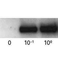
  • Precise tuning: precise control of protein level by controlling the dose of Shield1.
  • Reversible control: "protein on" to "protein off" for convincing gene-function studies.
  • What you get: each kit is supplied with a plasmid vector and an aliquot of Shield1.
  • NOTE: Most of the proteins that we tested show a better destabilization profile when the DD tag is fused to the N-terminus of the protein of interest (N Systems). Specific DD tag mutants for C-terminal tagging are available as well (C System); however, they have a slightly reduced destabilization activity in the absence of the Shield1 ligand.
  • For retrovirus production, use in combination with our Retro-X Universal Packaging System.
  • For lentivirus production, use in combination with our Lenti-X Packaging Single Shots.

More Information

Applications

  • Protein function in pathways
  • Functional analysis of subunits of a protein complex
  • Functional analysis of essential proteins

Additional product information

Please see the product's Certificate of Analysis for information about storage conditions, product components, and technical specifications. Please see the Kit Components List to determine kit components. Certificates of Analysis and Kit Components Lists are located under the Documents tab.


Control protein stability using a small molecule ligand, Shield1

Control protein stability with Shield1

ProteoTuner systems allow quick, predictable regulation of protein presence or absence in mammalian cells by acting directly on the protein, using a small, cell-permeable synthetic compound. When Shield1 is added to the medium, any fusion protein containing a DD domain can accumulate to detectable levels within 15–20 minutes. Conversely, upon Shield1 removal, the half-time for the protein's degradation can be as short as 30 minutes. This post-translational regulation offers many benefits, from speed and convenience to precise tuning and reversible control.

Technology explained Product selection guide

Takara Bio USA, Inc.
United States/Canada: +1.800.662.2566 • Asia Pacific: +1.650.919.7300 • Europe: +33.(0)1.3904.6880 • Japan: +81.(0)77.565.6999
FOR RESEARCH USE ONLY. NOT FOR USE IN DIAGNOSTIC PROCEDURES. © 2022 Takara Bio Inc. All Rights Reserved. All trademarks are the property of Takara Bio Inc. or its affiliate(s) in the U.S. and/or other countries or their respective owners. Certain trademarks may not be registered in all jurisdictions. Additional product, intellectual property, and restricted use information is available at takarabio.com.

Takara Bio USA, Inc. provides kits, reagents, instruments, and services that help researchers explore questions about gene discovery, regulation, and function. As a member of the Takara Bio Group, Takara Bio USA is part of a company that holds a leadership position in the global market and is committed to improving the human condition through biotechnology. Our mission is to develop high-quality innovative tools and services to accelerate discovery.

FOR RESEARCH USE ONLY. NOT FOR USE IN DIAGNOSTIC PROCEDURES (EXCEPT AS SPECIFICALLY NOTED).

Support
  • Contact us
  • Technical support
  • Customer service
  • Shipping & delivery
  • Sales
  • Feedback
Products
  • New products
  • Special offers
  • Instrument & reagent services
Learning centers
  • NGS
  • Gene function
  • Stem cell research
  • Protein research
  • PCR
  • Cloning
  • Nucleic acid purification
About
  • Our brands
  • Careers
  • Events
  • Blog
  • Need help?
  • Announcements
  • Quality and compliance
  • That's Good Science!
Facebook Twitter  LinkedIn

©2023 Takara Bio Inc. All Rights Reserved.

Region - North America Privacy Policy Terms and Conditions Terms of Use

Top


  • COVID-19 research
  • Viral detection with qPCR
  • SARS-CoV-2 pseudovirus
  • Human ACE2 stable cell line
  • Viral RNA isolation
  • Viral and host sequencing
  • Vaccine development
  • CRISPR screening
  • Drug discovery
  • Immune profiling
  • Publications
  • Next-generation sequencing
  • RNA-seq
  • DNA-seq
  • Single-cell NGS automation
  • Reproductive health
  • Bioinformatics tools
  • Whole genome amplification
  • Immune profiling
  • Diagnostic solutions
  • Reproductive health
  • Real-time PCR
  • Real-time PCR kits
  • Reverse transcription prior to qPCR
  • High-throughput qPCR solutions
  • RNA extraction and analysis for real-time qPCR
  • Stem cell research
  • Media and supplements
  • Stem cells and stem cell-derived cells
  • Single-cell cloning of edited hiPS cells
  • mRNA and cDNA synthesis
  • In vitro transcription
  • cDNA synthesis kits
  • Reverse transcriptases
  • RACE kits
  • Purified cDNA & genomic DNA
  • Purified total RNA and mRNA
  • PCR
  • Most popular polymerases
  • High-yield PCR
  • High-fidelity PCR
  • GC rich PCR
  • PCR master mixes
  • Cloning
  • In-Fusion seamless cloning
  • Competent cells
  • Ligation kits
  • Restriction enzymes
  • Nucleic acid purification
  • Plasmid purification kits
  • Genomic DNA purification kits
  • DNA cleanup kits
  • RNA purification kits
  • Cell-free DNA purification kits
  • Microbiome
  • Gene function
  • Gene editing
  • Viral transduction
  • Fluorescent proteins
  • T-cell transduction and culture
  • Tet-inducible expression systems
  • Transfection reagents
  • Cell biology assays
  • Protein research
  • Purification products
  • Two-hybrid and one-hybrid systems
  • Mass spectrometry reagents
  • Antibodies and ELISAs
  • Primary antibodies and ELISAs by research area
  • Fluorescent protein antibodies
  • New products
  • Special offers
  • Free samples
  • TB Green qPCR sale
  • PrimeSTAR enzyme promo
  • Try BcaBEST DNA Polymerase ver.2.0
  • RNA purification sale
  • Capturem IP and Co-IP sale
  • Baculovirus titration kits early access program
  • NGS bundle and save
  • Free sample: PrimePath Direct Saliva SARS-CoV-2 Detection Kit
  • TALON his-tag purification resin special offer
  • GoStix Plus special offers
  • PCR samples
  • OEM
  • Capabilities and installations
  • OEM enzyme FAQs
  • Enzyme samples for commerical assay developers
  • OEM process
  • Instrument services
  • Apollo services
  • ICELL8 services
  • SmartChip services
  • Stem cell services
  • Clinical-grade stem cell services
  • Research-grade stem cell services
  • Outsourcing stem cell-based disease model development
  • Gene and cell therapy manufacturing services
  • Services
  • Facilities
  • Our process
  • Resources
  • Customer service
  • Sales
  • Make an appointment with your sales rep
  • Shipping & delivery
  • Technical support
  • Feedback
  • Online tools
  • GoStix Plus FAQs
  • Partnering & Licensing
  • Vector information
  • Vector document overview
  • Vector document finder
Takara Bio's award-winning GMP-compliant manufacturing facility in Kusatsu, Shiga, Japan.

Partner with Takara Bio!

Takara Bio is proud to offer GMP-grade manufacturing capabilities at our award-winning facility in Kusatsu, Shiga, Japan.

  • Automation systems
  • SmartChip Real-Time PCR System introduction
  • ICELL8 introduction
  • Next-generation sequencing
  • Technical notes
  • Featured kits
  • Technology and application overviews
  • FAQs and tips
  • DNA-seq protocols
  • Bioinformatics resources
  • Webinars
  • cDNA synthesis
  • Real-time PCR
  • Overview
  • Reaction size guidelines
  • Guest webinar: extraction-free SARS-CoV-2 detection
  • Guest webinar: developing and validating molecular diagnostic tests
  • Technical notes
  • Nucleic acid purification
  • Nucleic acid extraction webinars
  • Product demonstration videos
  • Product finder
  • Plasmid kit selection guide
  • RNA purification kit finder
  • PCR
  • Citations
  • Selection guides
  • Technical notes
  • FAQ
  • Cloning
  • In-Fusion Cloning general information
  • Primer design and other tools
  • In‑Fusion Cloning tips and FAQs
  • Applications and technical notes
  • Stem cell research
  • Overview
  • Protocols
  • Technical notes
  • Gene function
  • Gene editing
  • Viral transduction
  • T-cell transduction and culture
  • Inducible systems
  • Cell biology assays
  • Protein research
  • Capturem technology
  • Antibody immunoprecipitation
  • His-tag purification
  • Other tag purification
  • Expression systems
  • Antibodies and ELISA
  • Molecular diagnostics
  • Interview: adapting to change with Takara Bio
  • Applications
  • Solutions
  • Partnering
  • Webinar: Speeding up diagnostic development
  • Contact us
  • Vaccine development
  • Characterizing the viral genome and host response
  • Identifying and cloning vaccine targets
  • Expressing and purifying vaccine targets
  • Immunizing mice and optimizing vaccine targets
  • Pathogen detection
  • Sample prep
  • Detection methods
  • Identification and characterization
  • SARS-CoV-2
  • Antibiotic-resistant bacteria
  • Food crop pathogens
  • Waterborne disease outbreaks
  • Viral-induced cancer
  • Immunotherapy research
  • T-cell therapy
  • Antibody therapeutics
  • T-cell receptor profiling
  • TBI initiatives in cancer therapy
  • Cancer research
  • Sample prep from FFPE tissue
  • Sample prep from plasma
  • Cancer biomarker discovery
  • Cancer biomarker quantification
  • Single cancer cell analysis
  • Cancer genomics and epigenomics
  • HLA typing in cancer
  • Gene editing for cancer therapy/drug discovery
  • Alzheimer's disease research
  • Antibody engineering
  • Sample prep from FFPE tissue
  • Single-cell sequencing
  • Reproductive health technologies
  • Preimplantation genetic testing
  • ESM Collection Kit forms
Create a web account with us

Log in to enjoy additional benefits

Want to save this information?

An account with takarabio.com entitles you to extra features such as:

•  Creating and saving shopping carts
•  Keeping a list of your products of interest
•  Saving all of your favorite pages on the site*
•  Accessing restricted content

*Save favorites by clicking the star () in the top right corner of each page while you're logged in.

Create an account to get started

  • BioView blog
  • Automation
  • Cancer research
  • Career spotlights
  • Current events
  • Customer stories
  • Gene editing
  • Research news
  • Single-cell analysis
  • Stem cell research
  • Tips and troubleshooting
  • Women in STEM
  • That's Good Support!
  • About our blog
  • That's Good Science!
  • SMART-Seq Pro Biomarker Discovery Contest
  • DNA extraction educational activity
  • That's Good Science Podcast
  • Season one
  • Season two
  • Season three
  • Our brands
  • Takara
  • Clontech
  • Cellartis
  • Our history
  • Announcements
  • Events
  • Biomarker discovery events
  • Calendar
  • Conferences
  • Speak with us
  • Careers
  • Company benefits
  • Trademarks
  • License statements
  • Quality statement
  • Takara Bio affiliates & distributors
  • United States and Canada
  • China
  • Japan
  • Korea
  • Europe
  • India
  • Affiliates & distributors, by country
  • Need help?
  • Privacy request
  • Website FAQs

That's GOOD Science!

What does it take to generate good science? Careful planning, dedicated researchers, and the right tools. At Takara Bio, we thoughtfully develop best-in-class products to tackle your most challenging research problems, and have an expert team of technical support professionals to help you along the way, all at superior value.

Explore what makes good science possible

 Customer Login
 View Cart (0)
  • Home
  • Products
  • Services & Support
  • Learning centers
  • APPLICATIONS
  • About
  • Contact Us
  •  Customer Login
  • Register
  •  View Cart (0)

Takara Bio USA, Inc. provides kits, reagents, instruments, and services that help researchers explore questions about gene discovery, regulation, and function. As a member of the Takara Bio Group, Takara Bio USA is part of a company that holds a leadership position in the global market and is committed to improving the human condition through biotechnology. Our mission is to develop high-quality innovative tools and services to accelerate discovery.

FOR RESEARCH USE ONLY. NOT FOR USE IN DIAGNOSTIC PROCEDURES (EXCEPT AS SPECIFICALLY NOTED).

Clontech, TaKaRa, cellartis

  • Products
  • COVID-19 research
  • Next-generation sequencing
  • Diagnostic solutions
  • Real-time PCR
  • Stem cell research
  • mRNA and cDNA synthesis
  • PCR
  • Cloning
  • Nucleic acid purification
  • Gene function
  • Protein research
  • Antibodies and ELISA
  • New products
  • Special offers
  • COVID-19 research
  • Viral detection with qPCR
  • SARS-CoV-2 pseudovirus
  • Human ACE2 stable cell line
  • Viral RNA isolation
  • Viral and host sequencing
  • Vaccine development
  • CRISPR screening
  • Drug discovery
  • Immune profiling
  • Publications
  • Next-generation sequencing
  • RNA-seq
  • DNA-seq
  • Single-cell NGS automation
  • Reproductive health
  • Bioinformatics tools
  • Whole genome amplification
  • Immune profiling
  • Diagnostic solutions
  • Reproductive health
  • Real-time PCR
  • Real-time PCR kits
  • Reverse transcription prior to qPCR
  • High-throughput qPCR solutions
  • RNA extraction and analysis for real-time qPCR
  • Stem cell research
  • Media and supplements
  • Stem cells and stem cell-derived cells
  • Single-cell cloning of edited hiPS cells
  • mRNA and cDNA synthesis
  • In vitro transcription
  • cDNA synthesis kits
  • Reverse transcriptases
  • RACE kits
  • Purified cDNA & genomic DNA
  • Purified total RNA and mRNA
  • PCR
  • Most popular polymerases
  • High-yield PCR
  • High-fidelity PCR
  • GC rich PCR
  • PCR master mixes
  • Cloning
  • In-Fusion seamless cloning
  • Competent cells
  • Ligation kits
  • Restriction enzymes
  • Nucleic acid purification
  • Plasmid purification kits
  • Genomic DNA purification kits
  • DNA cleanup kits
  • RNA purification kits
  • Cell-free DNA purification kits
  • Microbiome
  • Gene function
  • Gene editing
  • Viral transduction
  • Fluorescent proteins
  • T-cell transduction and culture
  • Tet-inducible expression systems
  • Transfection reagents
  • Cell biology assays
  • Protein research
  • Purification products
  • Two-hybrid and one-hybrid systems
  • Mass spectrometry reagents
  • Antibodies and ELISA
  • Primary antibodies and ELISAs by research area
  • Fluorescent protein antibodies
  • Special offers
  • Free samples
  • TB Green qPCR sale
  • PrimeSTAR enzyme promo
  • Try BcaBEST DNA Polymerase ver.2.0
  • RNA purification sale
  • Capturem IP and Co-IP sale
  • Baculovirus titration kits early access program
  • NGS bundle and save
  • Free sample: PrimePath Direct Saliva SARS-CoV-2 Detection Kit
  • TALON his-tag purification resin special offer
  • GoStix Plus special offers
  • PCR samples
  • Services & Support
  • OEM
  • Instrument services
  • Stem cell services
  • Gene and cell therapy manufacturing
  • Customer service
  • Sales
  • Shipping & delivery
  • Technical support
  • Feedback
  • Online tools
  • Partnering & Licensing
  • Vector information
  • OEM
  • Capabilities and installations
  • OEM enzyme FAQs
  • Enzyme samples for commerical assay developers
  • OEM process
  • Instrument services
  • Apollo services
  • ICELL8 services
  • SmartChip services
  • Stem cell services
  • Clinical-grade stem cell services
  • Research-grade stem cell services
  • Outsourcing stem cell-based disease model development
  • Gene and cell therapy manufacturing
  • Services
  • Facilities
  • Our process
  • Resources
  • Sales
  • Make an appointment with your sales rep
  • Online tools
  • GoStix Plus FAQs
  • Vector information
  • Vector document overview
  • Vector document finder
  • Learning centers
  • Automation systems
  • Next-generation sequencing
  • cDNA synthesis
  • Real-time PCR
  • Nucleic acid purification
  • PCR
  • Cloning
  • Stem cell research
  • Gene function
  • Protein research
  • Antibodies and ELISA
  • Automation systems
  • SmartChip Real-Time PCR System introduction
  • ICELL8 introduction
  • Next-generation sequencing
  • Technical notes
  • Featured kits
  • Technology and application overviews
  • FAQs and tips
  • DNA-seq protocols
  • Bioinformatics resources
  • Webinars
  • Real-time PCR
  • Overview
  • Reaction size guidelines
  • Guest webinar: extraction-free SARS-CoV-2 detection
  • Guest webinar: developing and validating molecular diagnostic tests
  • Technical notes
  • Nucleic acid purification
  • Nucleic acid extraction webinars
  • Product demonstration videos
  • Product finder
  • Plasmid kit selection guide
  • RNA purification kit finder
  • PCR
  • Citations
  • Selection guides
  • Technical notes
  • FAQ
  • Cloning
  • In-Fusion Cloning general information
  • Primer design and other tools
  • In‑Fusion Cloning tips and FAQs
  • Applications and technical notes
  • Stem cell research
  • Overview
  • Protocols
  • Technical notes
  • Gene function
  • Gene editing
  • Viral transduction
  • T-cell transduction and culture
  • Inducible systems
  • Cell biology assays
  • Protein research
  • Capturem technology
  • Antibody immunoprecipitation
  • His-tag purification
  • Other tag purification
  • Expression systems
  • APPLICATIONS
  • Molecular diagnostics
  • Vaccine development
  • Pathogen detection
  • Immunotherapy research
  • Cancer research
  • Alzheimer's disease research
  • Reproductive health technologies
  • Molecular diagnostics
  • Interview: adapting to change with Takara Bio
  • Applications
  • Solutions
  • Partnering
  • Webinar: Speeding up diagnostic development
  • Contact us
  • Vaccine development
  • Characterizing the viral genome and host response
  • Identifying and cloning vaccine targets
  • Expressing and purifying vaccine targets
  • Immunizing mice and optimizing vaccine targets
  • Pathogen detection
  • Sample prep
  • Detection methods
  • Identification and characterization
  • SARS-CoV-2
  • Antibiotic-resistant bacteria
  • Food crop pathogens
  • Waterborne disease outbreaks
  • Viral-induced cancer
  • Immunotherapy research
  • T-cell therapy
  • Antibody therapeutics
  • T-cell receptor profiling
  • TBI initiatives in cancer therapy
  • Cancer research
  • Sample prep from FFPE tissue
  • Sample prep from plasma
  • Cancer biomarker discovery
  • Cancer biomarker quantification
  • Single cancer cell analysis
  • Cancer genomics and epigenomics
  • HLA typing in cancer
  • Gene editing for cancer therapy/drug discovery
  • Alzheimer's disease research
  • Antibody engineering
  • Sample prep from FFPE tissue
  • Single-cell sequencing
  • Reproductive health technologies
  • Preimplantation genetic testing
  • ESM Collection Kit forms
  • About
  • BioView blog
  • That's Good Science!
  • Our brands
  • Our history
  • Announcements
  • Events
  • Careers
  • Trademarks
  • License statements
  • Quality and compliance
  • Takara Bio affiliates & distributors
  • Need help?
  • Website FAQs
  • BioView blog
  • Automation
  • Cancer research
  • Career spotlights
  • Current events
  • Customer stories
  • Gene editing
  • Research news
  • Single-cell analysis
  • Stem cell research
  • Tips and troubleshooting
  • Women in STEM
  • That's Good Support!
  • About our blog
  • That's Good Science!
  • SMART-Seq Pro Biomarker Discovery Contest
  • DNA extraction educational activity
  • That's Good Science Podcast
  • Season one
  • Season two
  • Season three
  • Our brands
  • Takara
  • Clontech
  • Cellartis
  • Events
  • Biomarker discovery events
  • Calendar
  • Conferences
  • Speak with us
  • Careers
  • Company benefits
  • Takara Bio affiliates & distributors
  • United States and Canada
  • China
  • Japan
  • Korea
  • Europe
  • India
  • Affiliates & distributors, by country
  • Need help?
  • Privacy request
  • Products
  • Services & Support
  • Learning centers
  • APPLICATIONS
  • About
  • Contact Us