

Trekker Single-Cell Spatial Mapping Kit resources
Introducing a new class of spatial technology
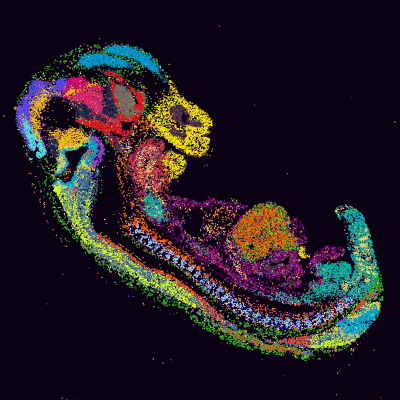
Acquire spatial and single-cell data in a single experiment, without sacrificing sensitivity or resolution.
- Analyze without deconvolution, segmentation, or complex algorithms to achieve true single-cell resolution
- Preserve the quality and sensitivity of your single-cell data with spatial barcodes that are donated to nuclei
- Gain single-cell spatial data by adding Trekker kits to your single-cell NGS workflow
Workflow
The one-hour Trekker workflow seamlessly integrates upstream of your single-cell assay.

The core of the Trekker technology lies in its spatially barcoded surface, composed of a bead monolayer. A 25 µm frozen tissue section is placed onto this barcoded substrate. Upon exposure to UV light, oligonucleotides carrying spatial barcodes are cleaved from the beads and attach to the nuclei in their vicinity. The tissue is then dissociated from the substrate, and the nuclei are isolated. Single-nucleus RNA sequencing (snRNA-seq) is performed on these isolated nuclei containing spatial barcode oligos. Spatial barcode oligos are captured and amplified alongside cellular RNA. For each sample, two sequencing libraries are generated—one for gene expression data and another for spatial barcodes. A custom analysis pipeline is used to map the position of each nucleus based on the spatial barcodes it contains. Integrating the Trekker protocol adds just one hour upstream of standard snRNA-seq workflows.
Webinar: Unlocking spatial multiomics with Trekker solutions
Topics covered:
- Adding spatial multiomics solutions to single-cell sequencing workflows
- Bringing spatial context to ATAC-seq and V(D)J sequencing alongside gene expression analysis
- Unlocking a deeper view of tissue organization and cellular interactions
Meet the presenter
Christina Chang, PhD
Dr. Christina Chang is the Director of Assay Applications, Spatial Genomics R&D at Takara Bio USA, Inc. She leads assay and applications development, driving the commercialization of novel spatial genomics products. Christina has extensive experience in single-cell multiomics technologies and has contributed to numerous inventions and commercial launches of single-cell and spatial genomics research products. Christina received her PhD in Immunology from the University of California, San Diego.

Trekker protocol videos
Watch how to generate spatially tagged, isolated single nuclei from fresh-frozen tissue samples.
Trekker FAQs
Learn more about using Trekker technology for single-cell spatial biology research.
Advancing discovery with spatial multiomics—accessible tools for high-resolution insights
Webinar series: Advancing discovery with spatial multiomics—accessible tools for high-resolution insights.
Takara Bio USA, Inc.
United States/Canada: +1.800.662.2566 • Asia Pacific: +1.650.919.7300 • Europe: +33.(0)1.3904.6880 • Japan: +81.(0)77.565.6999
FOR RESEARCH USE ONLY. NOT FOR USE IN DIAGNOSTIC PROCEDURES. © 2025 Takara Bio Inc. All Rights Reserved. All trademarks are the property of Takara Bio Inc. or its affiliate(s) in the U.S. and/or other countries or their respective owners. Certain trademarks may not be registered in all jurisdictions. Additional product, intellectual property, and restricted use information is available at takarabio.com.
