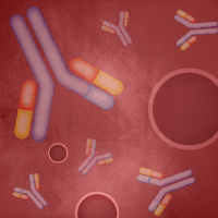
631076
Lenti-X™ Actin Dynamics Monitoring Kit
Each
Inquire for Quotation
*
Use this kit to monitor the highly dynamic behavior of the actin filament system in live cells. The Lenti-X Actin Dynamics Monitoring Kit includes lentiviral vectors encoding actin fusions to DD-AcGFP1 (green, destabilized) and mCherry (red), as well as the DD's stabilizing ligand Shield1. Once your cells have been infected with the included vectors, DD-AcGFP1-Actin is continuously targeted for degradation by the proteasomes, unless the cells are cultured in medium containing the stabilizing ligand Shield1. By contrast, mCherry-Actin (which does not contain the DD) has normal stability and is constitutively present in the cell.
Adding and removing Shield1 creates a "pulse-chase" like set of conditions which allow you to monitor how newly synthesized, Shield-stabilized DD-AcGFP1-Actin (green) is integrated into the existing (red) mCherry-Actin actin filament network.
Notice to purchaser
Our products are to be used for Research Use Only . They may not be used for any other purpose, including, but not limited to, use in humans, therapeutic or diagnostic use, or commercial use of any kind. Our products may not be transferred to third parties, resold, modified for resale, or used to manufacture commercial products or to provide a service to third parties without our prior written approval.
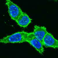
631078
pLVX-mCherry-Actin Vector
10 ug
Inquire for Quotation
*
This lentiviral expression vector encodes human β-actin fused to mCherry, an extremely bright red fluorescent protein. pLVX-mCherry-Actin constitutively expresses the mCherry-Actin fusion protein from P CMV IE when transduced into target cells. The mCherry-Actin fusion protein is incorporated into actin filaments, and allows for visualization of actin-containing subcellular structures in live cells as well as fixed cells. pLVX-mCherry-Actin is not designed to be used as a cloning vector.
To package the vector into high-titer, replication-incompetent lentiviral particles, we recommend using Lenti-X
Notice to purchaser
Our products are to be used for Research Use Only . They may not be used for any other purpose, including, but not limited to, use in humans, therapeutic or diagnostic use, or commercial use of any kind. Our products may not be transferred to third parties, resold, modified for resale, or used to manufacture commercial products or to provide a service to third parties without our prior written approval.
632408
pDsRed2-Nuc Vector
20 ug
USD $615.00
pDsRed2-Nuc encodes Discosoma sp. red fluorescent protein (DsRed2) fused with three copies of the nuclear localization signal (NLS) of the SV40 T-antigen. The NLS sequences are fused to the 3'-end of DsRed2. DsRed2 is a human codon-optimized variant of wild-type DsRed that has been engineered for faster maturation and lower non-specific aggregation. pDsRed2-Nuc is designed for fluorescent labeling of the nucleus in living cells.
Notice to purchaser
Our products are to be used for Research Use Only . They may not be used for any other purpose, including, but not limited to, use in humans, therapeutic or diagnostic use, or commercial use of any kind. Our products may not be transferred to third parties, resold, modified for resale, or used to manufacture commercial products or to provide a service to third parties without our prior written approval.
632409
pDsRed2-ER Vector
20 ug
USD $615.00
pDsRed2-ER is a mammalian expression vector designed to label the endoplasmic reticulum in living cells. The vector encodes a fusion consisting of Discosoma sp. red fluorescent protein (DsRed2); the endoplasmic reticulum (ER) targeting sequence of calreticulin, fused to the 5' end of DsRed2; and the ER retention sequence, KDEL, fused to the 3' end of DsRed2. DsRed2 is a human codon-optimized variant of wild-type DsRed that has been engineered for faster maturation and lower non-specific aggregation.
pDsRed2-ER can be introduced into mammalian cells using any standard transfection method. If required, stable transformants can be selected using G418.
Notice to purchaser
Our products are to be used for Research Use Only . They may not be used for any other purpose, including, but not limited to, use in humans, therapeutic or diagnostic use, or commercial use of any kind. Our products may not be transferred to third parties, resold, modified for resale, or used to manufacture commercial products or to provide a service to third parties without our prior written approval.
632418
pDsRed2-Peroxi Vector
20 ug
USD $615.00
pDsRed2-Peroxi encodes a fusion of Discosoma sp. red fluorescent protein (DsRed2) and the peroxisomal targeting signal 1 (PTS1). The PTS1 sequence is fused to the 3'-end of DsRed2 and encodes the tripeptide SKL, which targets the fusion protein to the matrix of peroxisomes. DsRed2 is human codon-optimized for high expression in mammalian cells. pDsRed2-Peroxi is designed for fluorescent labeling of peroxisomes.
Notice to purchaser
Our products are to be used for Research Use Only . They may not be used for any other purpose, including, but not limited to, use in humans, therapeutic or diagnostic use, or commercial use of any kind. Our products may not be transferred to third parties, resold, modified for resale, or used to manufacture commercial products or to provide a service to third parties without our prior written approval.
632419
pDsRed2-Bid Vector
20 ug
USD $615.00
pDsRed2-Bid encodes a fusion of Discosoma sp. red fluorescent protein (DsRed2) and Bid, a member of the Bcl-2 family. In healthy, non-apoptotic cells, Bid resides in the cytosol as soluble protein. Upon induction of apoptosis, Bid translocates to mitochondria. In cells expressing the Bid-DsRed2 fusion, the translocation can be detected by fluorescence microscopy.
Notice to purchaser
Our products are to be used for Research Use Only . They may not be used for any other purpose, including, but not limited to, use in humans, therapeutic or diagnostic use, or commercial use of any kind. Our products may not be transferred to third parties, resold, modified for resale, or used to manufacture commercial products or to provide a service to third parties without our prior written approval.
632421
pDsRed2-Mito Vector
20 ug
USD $615.00
pDsRed2-Mito encodes a fusion of Discosoma sp. red fluorescent protein (DsRed2) and a mitochondrial targeting sequence of human cytochrome c oxidase subunit VIII (Mito). The targeting sequence is fused to the 5'-end of DsRed2, which is human codon-optimized for high expression in mammalian cells. pDsRed2-Mito is designed for fluorescent labeling of mitochondria.
Notice to purchaser
Our products are to be used for Research Use Only . They may not be used for any other purpose, including, but not limited to, use in humans, therapeutic or diagnostic use, or commercial use of any kind. Our products may not be transferred to third parties, resold, modified for resale, or used to manufacture commercial products or to provide a service to third parties without our prior written approval.
632431
pAcGFP1-Nuc Vector
20 ug
Inquire for Quotation
*
pAcGFP1-Nuc encodes a humanized green fluorescent protein (GFP) derived from Aequorea coerulescens . The fluorescent protein is fused at its C-terminus to three copies of the nuclear localization signal (NLS) of the SV40 T-antigen. This vector is designed a marker for visualizing the nucleus in living or fixed cells by fluorescence microscopy.
Notice to purchaser
Our products are to be used for Research Use Only . They may not be used for any other purpose, including, but not limited to, use in humans, therapeutic or diagnostic use, or commercial use of any kind. Our products may not be transferred to third parties, resold, modified for resale, or used to manufacture commercial products or to provide a service to third parties without our prior written approval.
632432
pAcGFP1-Mito Vector
20 ug
Inquire for Quotation
*
pAcGFP1-Mito encodes a humanized green fluorescent protein (GFP) derived from Aequorea coerulescens . A mitochondrial targeting sequence from the precursor protein of human cytochrome c oxidase subunit VIII is fused to the N-terminus of the fluorescent protein. The fluorescence from pAcGFP1-Mito expression can be observed within the mitochondrial matrix inside the inner membrane. pAcGFP1-Mito can be used for specific labeling of mitochondria in living and fixed cells.
Notice to purchaser
Our products are to be used for Research Use Only . They may not be used for any other purpose, including, but not limited to, use in humans, therapeutic or diagnostic use, or commercial use of any kind. Our products may not be transferred to third parties, resold, modified for resale, or used to manufacture commercial products or to provide a service to third parties without our prior written approval.
632433
pHcRed1-Nuc Vector
20 ug
USD $615.00
pHcRed1-Nuc Vector encodes a far-red fluorescent protein HcRed1 fused with three copies of the nuclear localization signal (NLS) of the SV40 T-antigen. The NLS sequences are fused to the 3'-end of HcRed1. HcRed1 was generated by mutagenesis of a non-fluorescent chromoprotein from the reef coral Heteractis crispa . The coding sequence for HcRed1 is human codon-optimized for higher expression in mammalian cells. pHcRed1-Nuc is designed for visualizing the nucleus in living or fixed cells by fluorescence microscopy.
Notice to purchaser
Our products are to be used for Research Use Only . They may not be used for any other purpose, including, but not limited to, use in humans, therapeutic or diagnostic use, or commercial use of any kind. Our products may not be transferred to third parties, resold, modified for resale, or used to manufacture commercial products or to provide a service to third parties without our prior written approval.
632434
pHcRed1-Mito Vector
20 ug
USD $615.00
pHcRed1-Mito encodes a far-red fluorescent protein HcRed1 fused with mitochondrial targeting sequence from the precursor protein of human cytochrome c oxidase subunit VIII. HcRed1 is a fluorescent variant of a chromoprotein found in the reef coral Heteractis crispa . The coding sequence for HcRed1 has been human codon-optimized for higher expression in mammalian cells. pHcRed1-Mito can be used for specific labeling of mitochondria in living cells.
Notice to purchaser
Our products are to be used for Research Use Only . They may not be used for any other purpose, including, but not limited to, use in humans, therapeutic or diagnostic use, or commercial use of any kind. Our products may not be transferred to third parties, resold, modified for resale, or used to manufacture commercial products or to provide a service to third parties without our prior written approval.
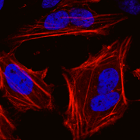
632453
pAcGFP1-Actin Vector
20 ug
Inquire for Quotation
*
pAcGFP1-Actin encodes a fusion protein consisting of Aequorea coerulescens green fluorescent protein (AcGFP1) fused at its C-terminus to the human cytoplasmic b-actin. The AcGFP1-Actin fusion protein incorporates into growing actin filaments, allowing visualization of the actin cytoskeleton in living or fixed cells.
Notice to purchaser
Our products are to be used for Research Use Only . They may not be used for any other purpose, including, but not limited to, use in humans, therapeutic or diagnostic use, or commercial use of any kind. Our products may not be transferred to third parties, resold, modified for resale, or used to manufacture commercial products or to provide a service to third parties without our prior written approval.
632464
pAcGFP1-Golgi Vector
20 ug
Inquire for Quotation
*
The pAcGFP1-Golgi Vector encodes a fusion protein consisting of Aequorea coerulescens green fluorescent protein (AcGFP1) fused at its N-terminus to 81 amino acids of the precursor to the human beta 1,4-galactosyltransferase (GT). AcGFP1 protein is optimized for brighter fluorescence and higher expression in mammalian cells. pAcGFP1-Golgi can be used for specific labeling of the trans-medial region of the Golgi apparatus in mammalian cells.
Notice to purchaser
Our products are to be used for Research Use Only . They may not be used for any other purpose, including, but not limited to, use in humans, therapeutic or diagnostic use, or commercial use of any kind. Our products may not be transferred to third parties, resold, modified for resale, or used to manufacture commercial products or to provide a service to third parties without our prior written approval.
632479
pDsRed-Monomer-Actin Vector
20 ug
Inquire for Quotation
*
pDsRed-Monomer-Actin encodes DsRed-Monomer, a monomeric mutant of the Discosoma sp. red fluorescent protein DsRed. In this vector, the DsRed-Monomer coding sequence is fused at its C-terminus to full-length human cytoplasmic ß-actin. The pDsRed-Monomer-Actin fusion protein incorporates into growing actin filaments, allowing visualization of the actin cytoskeleton in living or fixed cells.
Notice to purchaser
Our products are to be used for Research Use Only . They may not be used for any other purpose, including, but not limited to, use in humans, therapeutic or diagnostic use, or commercial use of any kind. Our products may not be transferred to third parties, resold, modified for resale, or used to manufacture commercial products or to provide a service to third parties without our prior written approval.
632480
pDsRed-Monomer-Golgi Vector
20 ug
Inquire for Quotation
*
pDsRed-Monomer-Golgi encodes DsRed-Monomer, a monomeric mutant of the Discosoma sp. red fluorescent protein DsRed. In this vector, the DsRed-Monomer coding sequence is fused at its N-terminus to the N-terminal 81 amino acids of human beta 1,4-galactosyltransferase (GT). The pDsRed-Monomer-Golgi fusion protein can be used for specific labeling of the trans-medial region of the Golgi apparatus in mammalian cells. A membrane anchoring signal peptide within the 81 amino acid sequence targets the fusion protein.
Notice to purchaser
Our products are to be used for Research Use Only . They may not be used for any other purpose, including, but not limited to, use in humans, therapeutic or diagnostic use, or commercial use of any kind. Our products may not be transferred to third parties, resold, modified for resale, or used to manufacture commercial products or to provide a service to third parties without our prior written approval.
632488
pAcGFP1-Tubulin Vector
20 ug
Inquire for Quotation
*
pAcGFP1-Tubulin encodes a fusion protein consisting of the AcGFP1 green fluorescent protein (GFP) and human a-tubulin. AcGFP1 is an Aequorea coerulescens GFP that has been optimized for brighter fluorescence and higher expression in mammalian cells. The AcGFP1-Tubulin fusion protein incorporates into growing microtubules, allowing visualization of the microtubule network in living or fixed cells.
Notice to purchaser
Our products are to be used for Research Use Only . They may not be used for any other purpose, including, but not limited to, use in humans, therapeutic or diagnostic use, or commercial use of any kind. Our products may not be transferred to third parties, resold, modified for resale, or used to manufacture commercial products or to provide a service to third parties without our prior written approval.
632490
pAcGFP1-Endo Vector
20 ug
Inquire for Quotation
*
pAcGFP1-Endo encodes a fusion protein consisting of the Aequorea coerulescens green fluorescent protein, c-Myc, and RhoB protein. The RhoB GTPase localizes the fusion protein to vesicles involved in endocytosis and allows monitoring of intracellular membrane traffic in living and fixed cells using fluorescence microscopy. The c-Myc epitope allows detection of the fusion protein using c-Myc antibodies. AcGFP1 is a variant of the Aequorea coerulescens green fluorescent protein (AcGFP) that has been optimized for brighter fluorescence and higher expression in mammalian cells.
Notice to purchaser
Our products are to be used for Research Use Only . They may not be used for any other purpose, including, but not limited to, use in humans, therapeutic or diagnostic use, or commercial use of any kind. Our products may not be transferred to third parties, resold, modified for resale, or used to manufacture commercial products or to provide a service to third parties without our prior written approval.
632491
pAcGFP1-Mem Vector
20 ug
Inquire for Quotation
*
pAcGFP1-Mem encodes a fusion protein consisting of the Aequorea coerulescens green fluorescent protein and the N-terminal membrane-targeting signal of neuromodulin (also called GAP-43). AcGFP1 is a variant of the Aequorea coerulescens green fluorescent protein (AcGFP) that has been optimized for brighter fluorescence and higher expression in mammalian cells. pAcGFP1-Mem can be used for labeling the plasma membrane.
Notice to purchaser
Our products are to be used for Research Use Only . They may not be used for any other purpose, including, but not limited to, use in humans, therapeutic or diagnostic use, or commercial use of any kind. Our products may not be transferred to third parties, resold, modified for resale, or used to manufacture commercial products or to provide a service to third parties without our prior written approval.
632493
pDsRed-Monomer-F Vector
20 ug
Inquire for Quotation
*
pDsRed-Monomer-F encodes a fusion protein consisting of the DsRed-Monomer red fluorescent protein and the farnesylation signal of c-Ha-ras. DsRed-Monomer is a monomeric mutant of the Discosoma sp. red fluorescent protein DsRed. The DsRed-Monomer coding sequence in this construct has been human codon-optimized for efficient expression and enhanced brightness in mammalian cells. pDsRed-Monomer-F can be used for labeling the inner face of the plasma membrane.
Notice to purchaser
Our products are to be used for Research Use Only . They may not be used for any other purpose, including, but not limited to, use in humans, therapeutic or diagnostic use, or commercial use of any kind. Our products may not be transferred to third parties, resold, modified for resale, or used to manufacture commercial products or to provide a service to third parties without our prior written approval.
632509
pAcGFP1-Mem Hyg Vector
20 ug
Inquire for Quotation
*
pAcGFP1-Mem Hyg encodes a fusion protein consisting of the Aequorea coerulescens green fluorescent protein and the N-terminal membrane-targeting signal of neuromodulin (also called GAP-43). AcGFP1 is a variant of the Aequorea coerulescens green fluorescent protein (AcGFP) that has been optimized for brighter fluorescence and higher expression in mammalian cells. pAcGFP1-Mem Hyg can be used for labeling the plasma membrane. The hygromycin resistance cassette allows you to create double-stable cell lines if this vector is used in conjunction with any other Living Colors vector that contains a neomycin resistance cassette.
Notice to purchaser
Our products are to be used for Research Use Only . They may not be used for any other purpose, including, but not limited to, use in humans, therapeutic or diagnostic use, or commercial use of any kind. Our products may not be transferred to third parties, resold, modified for resale, or used to manufacture commercial products or to provide a service to third parties without our prior written approval.
632510
pAcGFP1-F Hyg Vector
20 ug
Inquire for Quotation
*
pAcGFP1-F encodes a fusion protein consisting of the Aequorea coerulescens green fluorescent protein and the farnesylation signal of c-Ha-ras. AcGFP1 is a variant of the Aequorea coerulescens green fluorescent protein (AcGFP). The AcGFP1 coding sequence in this construct has been optimized for efficient expression and enhanced brightness in mammalian cells. pAcGFP1-F Hyg can be used for labeling the inner leaflet of the plasma membrane. The hygromycin resistance cassette allows you to create double-stable cell lines if this vector is used in conjunction with any other Living Colors vector that contains a neomycin resistance cassette.
Notice to purchaser
Our products are to be used for Research Use Only . They may not be used for any other purpose, including, but not limited to, use in humans, therapeutic or diagnostic use, or commercial use of any kind. Our products may not be transferred to third parties, resold, modified for resale, or used to manufacture commercial products or to provide a service to third parties without our prior written approval.
632511
pAcGFP1-F Vector
20 ug
Inquire for Quotation
*
pAcGFP1-F encodes a fusion protein consisting of the Aequorea coerulescens green fluorescent protein and the farnesylation signal of c-Ha-ras. AcGFP1 is a variant of the Aequorea coerulescens green fluorescent protein (AcGFP). The AcGFP1 coding sequence in this construct has been optimized for efficient expression and enhanced brightness in mammalian cells. pAcGFP1-F can be used for labeling the inner leaflet of the plasma membrane.
Notice to purchaser
Our products are to be used for Research Use Only . They may not be used for any other purpose, including, but not limited to, use in humans, therapeutic or diagnostic use, or commercial use of any kind. Our products may not be transferred to third parties, resold, modified for resale, or used to manufacture commercial products or to provide a service to third parties without our prior written approval.
632512
pDsRed-Monomer-Mem Vector
20 ug
Inquire for Quotation
*
pDsRed-Monomer-Mem encodes a fusion protein consisting of the DsRed-Monomer red fluorescent protein and the N-terminal membrane-targeting signal of neuromodulin (also called GAP-43). DsRed-Monomer is a monomeric mutant of the Discosoma sp. red fluorescent protein DsRed that has been optimized for efficient expression and enhanced brightness in mammalian cells. pDsRed-Monomer-Mem can be used for labeling the plasma membrane.
Notice to purchaser
Our products are to be used for Research Use Only . They may not be used for any other purpose, including, but not limited to, use in humans, therapeutic or diagnostic use, or commercial use of any kind. Our products may not be transferred to third parties, resold, modified for resale, or used to manufacture commercial products or to provide a service to third parties without our prior written approval.
632513
pDsRed-Monomer-Mem Hyg Vector
20 ug
Inquire for Quotation
*
pDsRed-Monomer-Mem Hyg encodes a fusion protein consisting of the DsRed-Monomer red fluorescent protein and the N-terminal membrane-targeting signal of neuromodulin (also called GAP-43). DsRed-Monomer is a monomeric mutant of the Discosoma sp. red fluorescent protein DsRed that has been optimized for efficient expression and enhanced brightness in mammalian cells. pDsRed-Monomer-Mem Hyg can be used for labeling the plasma membrane. The hygromycin resistance cassette allows you to create double-stable cell lines if this vector is used in conjunction with any other Living Colors vector that contains a neomycin resistance cassette.
Notice to purchaser
Our products are to be used for Research Use Only . They may not be used for any other purpose, including, but not limited to, use in humans, therapeutic or diagnostic use, or commercial use of any kind. Our products may not be transferred to third parties, resold, modified for resale, or used to manufacture commercial products or to provide a service to third parties without our prior written approval.
632514
pDsRed-Monomer-F Hyg Vector
20 ug
Inquire for Quotation
*
pDsRed-Monomer-F Hyg encodes a fusion protein consisting of the DsRed-Monomer red fluorescent protein and the farnesylation signal of c-Ha-ras. DsRed-Monomer is a monomeric mutant of the Discosoma sp. red fluorescent protein DsRed. The DsRed-Monomer coding sequence in this construct has been optimized for efficient expression and enhanced brightness in mammalian cells. pDsRed-Monomer-F Hyg can be used for labeling the inner leaflet of the plasma membrane. The hygromycin resistance cassette allows you to create double-stable cell lines if this vector is used in conjunction with any other Living Colors vector that contains a neomycin resistance cassette.
Notice to purchaser
Our products are to be used for Research Use Only . They may not be used for any other purpose, including, but not limited to, use in humans, therapeutic or diagnostic use, or commercial use of any kind. Our products may not be transferred to third parties, resold, modified for resale, or used to manufacture commercial products or to provide a service to third parties without our prior written approval.
632515
pIRES2-AcGFP1-Nuc Vector
20 ug
Inquire for Quotation
*
pIRES2-AcGFP1-Nuc encodes AcGFP1 with three identical copies of the nuclear localization signal (NLS) of the simian virus 40 large T antigen fused in tandem to its C-terminus. The gene encoding the AcGFP1-Nuc fusion protein is located downstream of the IRES2 sequence. This permits both the gene of interest (cloned into the MCS upstream of the IRES2 sequence) and the AcGFP1-Nuc gene to be translated from a single bicistronic mRNA. AcGFP-1-Nuc localizes to the nucleus of a transfected cell, offering an advantage over random localization in the cytosol. It is most useful for those who are interested in monitoring translocation events in and out of the nucleus.
Notice to purchaser
Our products are to be used for Research Use Only . They may not be used for any other purpose, including, but not limited to, use in humans, therapeutic or diagnostic use, or commercial use of any kind. Our products may not be transferred to third parties, resold, modified for resale, or used to manufacture commercial products or to provide a service to third parties without our prior written approval.
632583
pAutophagSENSE Vector
Each
Inquire for Quotation
*
The pAutophagSENSE vector expresses the mutant green fluorescent protein AcGFP1 (excitation maximum = 475 nm; emission maximum = 505 nm) fused to the mouse LC3 protein in mammalian cells. The fusion protein is incorporated into the autophagosomes and is used to monitor autophagosome formation in live cells. This process is monitored by following the redistribution of a green fluorescent fusion protein from the cytosol to the forming autophagosomes.
Notice to purchaser
Our products are to be used for Research Use Only . They may not be used for any other purpose, including, but not limited to, use in humans, therapeutic or diagnostic use, or commercial use of any kind. Our products may not be transferred to third parties, resold, modified for resale, or used to manufacture commercial products or to provide a service to third parties without our prior written approval.
632588
pPAmCherry-Mem Vector
10 ug
Inquire for Quotation
*
pPAmCherry-Mem Vector is mammalian expression vector encoding a fusion protein of PAmCherry and the N-terminal 20 amino acids of neuromodulin (GAP-43). PAmCherry is a photoactivatable mutant of the fluorescent protein mCherry. PAmCherry is non-fluorescence until photoactivated by a short exposure to light at a wavelength between 350 nm and 400 nm. The excitation/emission wavelengths of photoactivated PAmCherry are 564 nm and 595 nm. The GAP-43 fragment contains a signal for posttranslational palmitoylation of cysteins 3 and 4 that targets the fusion protein to the plasma membrane.
Notice to purchaser
Our products are to be used for Research Use Only . They may not be used for any other purpose, including, but not limited to, use in humans, therapeutic or diagnostic use, or commercial use of any kind. Our products may not be transferred to third parties, resold, modified for resale, or used to manufacture commercial products or to provide a service to third parties without our prior written approval.
632589
pPAmCherry-Actin Vector
10 ug
Inquire for Quotation
*
pPAmCherry-Actin Vector is a mammalian expression vector encoding PAmCherry, a photoactivatable mutant of the fluorescent protein mCherry, fused to human cytoplasmic beta-actin. PAmCherry is non-fluorescence until photoactivated by a short exposure to light at a wavelength between 350 nm and 400 nm. The excitation/emission wavelengths of photoactivated PAmCherry are 564 nm and 595 nm.
Notice to purchaser
Our products are to be used for Research Use Only . They may not be used for any other purpose, including, but not limited to, use in humans, therapeutic or diagnostic use, or commercial use of any kind. Our products may not be transferred to third parties, resold, modified for resale, or used to manufacture commercial products or to provide a service to third parties without our prior written approval.
632590
pPAmCherry-Tubulin Vector
10 ug
Inquire for Quotation
*
pPAmCherry-Tubulin Vector is a mammalian expression vector encoding PAmCherry, a photoactivatable mutant of the fluorescent protein mCherry, fused to human alpha-tubulin. PAmCherry is non-fluorescence until photoactivated by a short exposure to light at a wavelength between 350 nm and 400 nm. The excitation/emission wavelengths of photoactivated PAmCherry are 564 nm and 595 nm.
Notice to purchaser
Our products are to be used for Research Use Only . They may not be used for any other purpose, including, but not limited to, use in humans, therapeutic or diagnostic use, or commercial use of any kind. Our products may not be transferred to third parties, resold, modified for resale, or used to manufacture commercial products or to provide a service to third parties without our prior written approval.
632591
pPAmCherry-Mito Vector
10 ug
Inquire for Quotation
*
pPAmCherry-Mito Vector is a mammalian expression vector encoding PAmCherry, a photoactivatable mutant of the fluorescent protein mCherry, fused to the mitochondrial targeting sequence derived from the precursor subunit VIII of human cytochrome C oxidase. PAmCherry is non-fluorescence until photoactivated by a short exposure to light at a wavelength between 350 nm and 400 nm. The excitation/emission wavelengths of photoactivated PAmCherry are 564 nm and 595 nm.
Notice to purchaser
Our products are to be used for Research Use Only . They may not be used for any other purpose, including, but not limited to, use in humans, therapeutic or diagnostic use, or commercial use of any kind. Our products may not be transferred to third parties, resold, modified for resale, or used to manufacture commercial products or to provide a service to third parties without our prior written approval.