Small but mighty: gaining insight into pulmonary arterial hypertension with purified exosomes

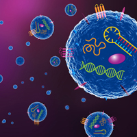
Despite their small size, extracellular vesicles (EVs), such as exosomes, play important roles in normal physiological processes (e.g., immune response, neuronal function, and stem cell maintenance) and disease pathology (e.g., cancer and liver disease). The secretion and uptake of EVs are critical steps in intercellular communication, facilitating the delivery of various cargos, including functional proteins and RNA molecules. A breakdown in this communication may lead to various disease states, making EV research of growing interest.
With the potential to reveal such key insights into human health and development, researchers have been looking for methods to work with EVs that don’t involve the traditional process of ultracentrifugation. While this has historically been the most commonly used method, it is time-consuming, suffers from low yield, requires specialized equipment, and may cause damage to the EVs themselves.
In a recent study, researchers at Stanford University Medical Center tackled this issue while delving into the underlying causes of pulmonary arterial hypertension (PAH), a serious condition that displays high blood pressure in vessels in the lungs. In their investigations, Yuan et al. discovered that one missing piece of the puzzle could be revealed by looking at exosomes from the cells that surround the affected vessels.
A broken repair mechanism
Pericytes are specialized mural cells that surround capillaries throughout the body and are crucial for the correct function and maturation of vasculature in all human tissue. The researchers here noted that reduced pericyte coverage of microvessels in the lungs is part of the pathology of PAH, and they decided to look into the mechanisms related to this small vessel loss, as it relates to pulmonary pericytes.
Healthy and PAH pulmonary microvascular endothelial cells (PMVECs) were tested for their ability to recruit pericytes—a necessary process for stabilizing new microvessels. Coculture of healthy lung pericytes with healthy PMVECs resulted in significant vascular network development, whereas PAH PMVECs under the same conditions failed to recruit healthy pericytes and showed no such vascular development.
The researchers looked to the Wnt/planar cell polarity (PCP) signaling pathway, as their own previous work had indicated that it has an important role in recruiting pericytes to PMVECs. Expression of well-characterized Wnt ligands was examined in healthy and PAH PMVECs. Healthy PMVECs showed a significant increase in the expression of Wnt5a when in coculture with pericytes, as compared to when they were seeded alone. PAH PMVECs did not exhibit this response, a fact that was also shown in Western blots of lysates from the cultures. A subsequent siRNA knockdown of Wnt5a in healthy PMVECs resulted in behavior similar to that of PAH PMVECs.
A failure to communicate
Wnts can have long-range effects, as they can move between cells as exosome cargo and remain biologically active (Luga et al. 2012; Zhang et al. 2014). Yuan et al. also saw evidence of exosomal packaging when staining PMVECs for Wnt5a. In healthy PMVECs, a diffuse staining pattern was seen when seeded alone, but organized, dense particles heavily clustered at the membrane were seen when cocultured with pericytes. While PAH PMVECs showed low Wnt5a expression in general, they additionally saw no significant change in organization or distribution when in coculture. With these results in hand, the researchers looked to isolate exosomes for further analysis of Wnt5a levels in PMVECs.
As mentioned earlier, ultracentrifugation is the most common method used for exosome isolation but is not without its problems. Here, the researchers turned to their newly developed ExoTIC technology that utilizes filter membranes to isolate exosomes from biofluids, allowing them to achieve better yields with less starting material. The resulting purified exosomes were used to treat PMVECs in monoculture and coculture. Additionally, Capturem EV spin columns were also used to isolate exosomes, and TEM images were taken to visually confirm the presence of exosomes in these eluates (Figure 1).

Figure 1. TEM image of a single exosome isolated from coculture using the Capturem Extracellular Vesicle Isolation Kit. Image kindly provided by Ke Yuan, PhD, as a researcher at Stanford University Medical Center (currently an assistant professor at Boston Children's Hospital).
When exosomes purified with the ExoTIC method were applied to monoculture and coculture with pericytes, the same pattern seen earlier emerged yet again—with healthy PMVECs showing an increase in exosomes and Wnt5a expression (as measured by Western blot and IF staining) when in the presence of pericytes, and PAH PMVECs showing no change.
Further testing was performed to see if these Wnt5a-containing exosomes would demonstrate expected biological activity. Indeed, when whole, purified exosomes from healthy PMVECs were added to cocultures of PAH PMVECs, pericyte recruitment was significantly increased. When repeating this protocol with whole exosomes purified from the aforementioned Wnt5a knockdown, a decrease in pericyte recruitment was observed.
Taken together, these data point to the delivery of Wnt5a via exosomes as key to the healthy behavior of the cells. When this cell-to-cell communication breaks down, so does the work needed to maintain and repair healthy blood vessels.
Good things come in small packages
Perhaps it’s unsurprising at this point, but small players often turn out to hold critical places in the larger picture of human health. Pericytes act to protect and support microvessels in the lungs, but if microvessels are damaged and unrepaired, life-threatening and progressive hypertension results. Exosomes, though small in size, are known to play important roles in cell signaling, and the pathology of PAH seems to be no exception. Conducting research on these submicron particles, without running into the bottlenecks of traditional methods, will surely prove useful in a wide range of studies. Our own R&D team has seen the benefits of alternative methods to ultracentrifugation for EV isolation, and we look forward to seeing where further innovations in EV research will lead scientists in their quests to understand and improve human health.
References
Luga, V., et al. Exosomes mediate stromal mobilization of autocrine Wnt-PCP signaling in breast cancer cell migration. Cell 151, 1542–1556 (2012).
Zhang, L. and Wrana, J. L. The emerging role of exosomes in Wnt secretion and transport. Curr. Opin. Genet. Dev. 27, 14–19 (2014).
Yuan, K., et al. Loss of endothelium-derived Wnt5a is associated with reduced pericyte recruitment and small vessel loss in pulmonary arterial hypertension. Circulation 139, 1710–1724 (2019).

Outperform ultracentrifugation with easy isolation of pure, concentrated extracellular vesicles
The Capturem extracellular (EV) vesicle isolation kits consistently generate pure, concentrated EV samples with enough yield for subsequent proteomic, genomic, and transcriptomic analyses. This unique protocol enables isolation of EVs from biofluids like plasma, serum, urine, milk, saliva, cell-conditioned media, and cerebrospinal fluid (CSF) with a simple workflow that takes less than 30 minutes.
Takara Bio USA, Inc.
United States/Canada: +1.800.662.2566 • Asia Pacific: +1.650.919.7300 • Europe: +33.(0)1.3904.6880 • Japan: +81.(0)77.565.6999
FOR RESEARCH USE ONLY. NOT FOR USE IN DIAGNOSTIC PROCEDURES. © 2025 Takara Bio Inc. All Rights Reserved. All trademarks are the property of Takara Bio Inc. or its affiliate(s) in the U.S. and/or other countries or their respective owners. Certain trademarks may not be registered in all jurisdictions. Additional product, intellectual property, and restricted use information is available at takarabio.com.


