pHEK293 Ultra Expression Vectors overview
- Ultra-high expression of a protein of interest in HEK 293 cells
- Flexibility of a single- or double-vector system
- Protein expression up to 10 times higher than with CMV-derived promoters
- Ideal for production of biomedically relevant proteins, including monoclonal antibodies
E. coli is commonly used to express recombinant proteins in life science research. However, mammalian cell-based expression systems have several advantages over E. coli, especially for eukaryotic protein expression. Expression in mammalian cells allows for proper protein folding and post-translational modification—features that are key for proper protein function in vitro. pHEK293 Ultra Expression Vectors enable ultra-high, transient overexpression of a protein of interest in human HEK 293 cells. Please visit our tech note for data specific to the use of these vectors for producing monoclonal antibodies.
Principles of high expression with the TAR-Tat system
High levels of recombinant protein expression with pHEK293 Ultra Expression Vectors is accomplished by a transcriptional activation mechanism based on the HIV-1 virus TAR-Tat system. The TAR (transactivation response) element is an RNA sequence derived from HIV-1 that forms a stem loop structure at the 5' end of the viral RNA. Tat is an RNA-binding protein that binds to TAR and promotes transcription.
pHEK293 Ultra Expression Vector I and Vector II systems incorporate the TAR-Tat elements in order to provide ultra-high expression of your protein of interest. Both Vector I and Vector II systems incorporate the TAR sequence in the 5' untranslated region of the target gene, where it will not affect the amino acid sequence of the expressed target protein. For the Vector I system, a Tat cassette is included on the same vector; the Vector II system includes the Tat cassette on a separate Enhancer Vector, enabling greater control of the protein of interest's expression level. With both systems, expression of the Tat protein greatly increases the expression of the protein of interest.
- pHEK293 Ultra Expression Vector I (Cat. # 3390)—Obtain high expression with the convenience of a single-plasmid system
- pHEK293 Ultra Expression Vector II (Cat. # 3392)—Optimize expression levels by varying the ratio of Vector II and the included Enhancer Vector

Schematic of protein overexpression using pHEK293 Ultra Expression Vectors. The TAR element forms a stem loop structure at the 5' end of the transcribed RNA. Expression of the Tat element produces the Tat protein which binds to the TAR element in the RNA loop structure. This activates TAR, which in turn promotes high expression of your protein of interest. The cassette in the image above is from the single-vector system, where TAR and Tat are both on the same plasmid as your target gene, enabling more convenient transfection. In the two-vector system, Tat is on a separate plasmid, allowing you to have control over expression levels by varying the ratio of the two vectors.
Visualization of high expression using fluorescent proteins
In order to assess protein expression capabilities, both pHEK293 Ultra Expression Vector systems were used to introduce a fluorescent protein (AcGFP1) into adherent and suspension HEK 293 cells. For comparison, the same assay was conducted with a vector expressing AcGFP1 from a CMV promoter (pBApo-CMV DNA) and a high-expression vector from Company A. Constructs were made by inserting AcGFP1 into the appropriate vectors (details listed in the table at the end of this page).
Higher fluorescence intensity was observed for the pHEK293 systems (conditions 2, 3, and 4) than for either of the other two vectors (conditions 1 and 5). Additionally, a color change in suspension cells was readily apparent to the naked eye for cells transfected with the pHEK293 vectors.

Visualization of AcGFP1 expression in adherent and suspension HEK 293 cells. Adherent cells (experimental conditions 1–5) were cultured in 12-well plates. Suspension cells (experimental conditions 6–10) were cultured in 125-ml flasks. Imaging was done two days post-transfection. Panel A. Fluorescent microscopy images (adherent cells). Panel B. Representative images of suspension cell cultures (condition 7 on the left; condition 10 on the right; images for conditions 6, 8, and 9 not shown, however, quantitative data for all conditions are shown in the figure below). For Panels A and B, experimental conditions are as follows. Conditions 1 and 6: pBApo-CMV; conditions 2 and 7: pHEK293 Ultra Expression Vector I; conditions 3 and 8: pHEK293 Ultra Expression Vector II + Enhancer Vector; conditions 4 and 9: same as 3 and 8, but with a larger amount of Enhancer Vector; conditions 5 and 10: Company A's high-expression vector.
Flow cytometry was used to quantify the expression levels of AcGFP1 in adherent and suspension cells. Cells transfected with pHEK293 Ultra Expression Vector I (conditions 2 and 7) and pHEK293 Ultra Expression Vector II (conditions 3, 4, 8, and 9) displayed fluorescence intensities 5–11 times higher than that obtained with a typical CMV promoter (conditions 1 and 6), and 2–7 times higher than Company A's high-expression vector (conditions 5 and 10). Additionally, cells transfected with the two-vector system displayed an expected increase in AcGFP1 expression when the amount of Enhancer Vector was raised from 0.008 µg (condition 3) to 0.2 µg (condition 4) for adherent cells, and from 0.24 µg (condition 8) to 6 µg (condition 9) for suspension cells.

Flow cytometry analysis of fluorescence intensity of AcGFP1 positive cells. AcGFP1 expression in adherent and suspension cells (transfected with constructs as described above) was quantified two days post-transfection. The table column directly below each bar on the graph lists the amount of each vector used in that specific experimental condition.
Ultra-high expression of recombinant proteins
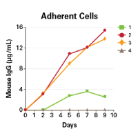
High expression of functional OKT3 monoclonal antibodies in mammalian cells
pHEK293 Ultra Expression Vectors provide ultra-high expression of recombinant proteins, including antibodies, in mammalian cells.
Takara Bio USA, Inc.
United States/Canada: +1.800.662.2566 • Asia Pacific: +1.650.919.7300 • Europe: +33.(0)1.3904.6880 • Japan: +81.(0)77.565.6999
FOR RESEARCH USE ONLY. NOT FOR USE IN DIAGNOSTIC PROCEDURES. © 2025 Takara Bio Inc. All Rights Reserved. All trademarks are the property of Takara Bio Inc. or its affiliate(s) in the U.S. and/or other countries or their respective owners. Certain trademarks may not be registered in all jurisdictions. Additional product, intellectual property, and restricted use information is available at takarabio.com.


