We use cookies to improve your browsing experience and provide meaningful content. Read our cookie policy. Accept
  •  Customer Login
  • Register
  •  View Cart (0)
  •  Customer Login
  • Register
  •  View Cart (0)

Takara Bio
  • Products
  • Services & Support
  • Learning centers
  • APPLICATIONS
  • About
  • Contact Us

Clontech Takara Cellartis

Close

  • ‹ Back to Antibody immunoprecipitation
  • IP and Co-IP of cardiac voltage-gated ion channel proteins
  • Tech note: thiophilic antibody purification resins
  • Tech note: IP and Co-IP
Capturem IP & Co-IP Kit Capturem IP & Co-IP Kit
antibody purification Antibody purification kits
Home › Learning centers › Protein research › Antibody immunoprecipitation › Tech note: IP and Co-IP

Protein research

  • Capturem technology
    • Capturem protocols
    • Capturem tech notes and applications
    • FAQs about Capturem technology
    • Capturem technology citations
    • Capturem posters
  • Antibody immunoprecipitation
    • IP and Co-IP of cardiac voltage-gated ion channel proteins
    • Tech note: thiophilic antibody purification resins
    • Tech note: IP and Co-IP
  • His-tag purification
    • Purification methods overview
    • TALON resin selection guide
    • Selection guide: His60 resin
    • xTractor Buffer is optimized for superior protein yield
    • Why tag a protein?
    • Tech note: cobalt resin
    • Simplified purification of active, secreted his-tagged proteins
    • Overview: His60
    • Tech note: Capturem technology
    • Tech note: Capturem large volume
    • Magnetic beads
    • FAQs: TALON
    • Protocols
      • Video: Capturem his maxiprep
      • Video: Capturem his miniprep
      • Visual protocol: Capturem his maxiprep
      • Visual protocol: Capturem his miniprep
      • Capturem nickel column reagent compatibility
      • TALON reagent compatibility
      • His60 reagent compatibility
      • TALON: Native vs denaturing purification
      • Protocol: denaturing purification with TALON resin, imidazole elution
      • Protocol: native purification with TALON resin, imidazole elution
      • Protocol: native purification with TALON resin, pH elution
  • Other tag purification
    • Streptavidin-based enrichment using Capturem technology
    • Selection guide: peptide tags
    • Myc-tagged protein purification overview
    • GST-tagged protein purification overview
  • Phosphoprotein and glycoprotein purification
    • Non-tagged protein purification overview
    • Phosphoprotein purification overview
  • Matchmaker Gold yeast two-hybrid systems
    • Matchmaker Gold Yeast Two-Hybrid System
    • Make your own library for yeast two-hybrid screening
    • Mate and Plate yeast two-hybrid cDNA libraries
    • Aureobasidin A for improved selectable drug resistance in yeast
  • Expression systems
    • Protein expression overview
    • Insect expression overview
    • Mammalian expression overview
    • pHEK293 Ultra expression overview
    • OKT3 expression in mammalian cells
    • Bacterial expression overview
New products
Need help?
Contact Sales
Capturem IP & Co-IP Kit Capturem IP & Co-IP Kit
antibody purification Antibody purification kits
Tech Note

Fast, efficient, and specific immunoprecipitation and co-immunoprecipitation with Capturem Protein A technology

  • Rapid Protein A column IP of PP2A B subunit
  • Compatibility with a variety of IP buffers
  • p53 and SV40 T Co-IP with Capturem Protein A technology
Introduction Results Conclusions Methods

Introduction  

For nearly half a century, immunoprecipitation (IP) has been a cornerstone of cell and molecular biology research. In addition, it has proven to be a valuable screening technique for the development of novel biologics, such as antibody-based therapeutics. As academic and biopharmaceutical researchers look to accelerate the application of bench discoveries to the clinic, there is a constant need for faster and more efficient immunoprecipitation protocols.

Capturem Protein A columns combine the specific antibody-binding properties of Protein A with novel Capturem technology to provide high-capacity, membrane-based affinity purification. The antibody-binding properties also make these columns ideal for use in IP and co-immunoprecipitation (Co-IP) experiments. After optimization of the purification parameters, we combined the Capturem Protein A columns with an optimized IP buffer set to create the Capturem IP & Co-IP Kit, which provides a complete, easy-to-use IP/Co-IP solution. The kit also has added utility for the purification and screening of antibody-based therapeutics, including antibody-drug conjugates. Furthermore, the benefits of this resin-free system—including speed, ease-of-use, flexibility, and yield—make it a powerful tool for both academic research institutions and biopharmaceutical organizations.

Results  

Rapid Protein A column immunoprecipitation

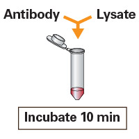
The same no-waiting workflow that provides fast, high-quality antibody purification also enables immunoprecipitation experiments to be completed quickly and easily. Following a 10-minute incubation of the antibody-antigen complex, the kit requires only five minutes of hands-on time for efficient IP. In the experiment below, NIH3T3 lysates were incubated with 1 µg of a protein phosphatase type 2A (PP2A) B subunit antibody before being applied to equilibrated Capturem Protein A spin columns. After washing with 300 µl of wash buffer and eluting in 100 µl of elution buffer, the various fractions were resolved by gel electrophoresis, transferred onto polyvinylidene fluoride (PVDF) membranes, and probed with an anti-PP2A B antibody. The Capturem Protein A spin columns yielded excellent results, with a strong protein PP2A B signal in the eluate fraction. Note: This data was produced using buffer formulations that differ from those found in the final Capturem IP & Co-IP Kit (see Methods: Immunoprecipitation performed with Capturem Protein A columns).

IP of PP2A B with Capturem Protein A columns

Figure 1. Immunoprecipitation performed with Capturem Protein A columns. NIH3T3 cell lysates were incubated with 1 µg of PP2A B subunit antibody for 10 min. The antibody-lysate complex was applied to equilibrated Protein A spin columns. Following centrifugation and washing steps, elution was carried out in 100 µl of elution buffer. Fractions were resolved by gel electrophoresis, transferred onto PVDF membranes, and probed with an anti-PP2A B antibody. The protein in the eluate fraction corresponds to the PP2A B subunit (52 kDa).

Compatibility with a variety of immunoprecipitation buffers

Capturem Protein A products provide an excellent level of flexibility to accommodate a wide range of experimental conditions, including those performed with IP buffers from other kits. To test this aspect of performance, NIH3T3 cell lysates were incubated with a PP2A B subunit antibody in buffers provided with either IP Kit A, IP Kit B, or standalone lysis buffer C. After incubation, the antibody-lysate mixture was loaded onto Capturem Protein A spin columns that had been equilibrated with the corresponding IP buffer. Once centrifugation and wash steps were complete, elution was performed in 30 µl of elution buffer appropriate for each sample set (Figure 2; Capturem Protein A columns A1, B1, C1). Elution was repeated to ensure complete recovery of antibody complexes (Figure 2; Capturem Protein A columns A2, B2, C2). IP was also performed using the full IP kits A and B, per their respective protocols (Figure 2; IP Kit A, IP Kit B), starting with the same amount of lysate and antibody as used for Capturem columns.

All samples were resolved on a gel, transferred to PVDF membranes, and probed for the PP2A B subunit using an appropriate antibody. The gel below shows the presence of the subunit in the original sample (OS), which is greatly enriched in the immunoprecipitated samples. Results from the Capturem Protein A columns showed strong PP2A B protein signal in the first elution, regardless of the IP buffer used, with the highest elution level for the Capturem samples eluted with a low pH glycine buffer (A1, C1). Note: This data was also produced using buffer formulations that differ from those found in the final Capturem IP & Co-IP Kit (see Methods: Immunoprecipitation performed with Capturem Protein A columns).

Figure 2. Capturem Protein A columns are compatible with a variety of buffers.NIH3T3 cell lysates (100 µl, containing 130 µg of total protein) were incubated with 0.6 µg of protein phosphatase type 2A B subunit antibody in the IP buffers provided with either IP Kit A or B, or lysis buffer C, bringing the total volume up to 400 µl. The antibody-lysate mixture was incubated at 4°C for 1 hour and then loaded on Capturem Protein A columns that had been equilibrated with the corresponding IP buffer. Bound immunocomplexes were then eluted twice with 30 µl of an appropriate elution buffer. IPs were also done using the full A or B IP kits. All elution samples were then resolved on a gel, transferred to PVDF membranes, and probed for the PP2A B subunit using an appropriate antibody. The presence of the PP2A B subunit in the original sample is shown in lane OS. Elution samples #1 and #2 are shown for those performed entirely with IP Kit A and IP Kit B. Elution samples #1 and #2 for Capturem Protein A columns are shown for those performed with buffers from IP Kit A (A1, A2), IP Kit B (B1, B2), and the standalone lysis buffer (C1, C2).

Co-immunoprecipitation with Capturem Protein A Minipreps

p53 is a 53-kDa nuclear phosphoprotein that functions as a tumor suppressor and is involved in inhibiting cell proliferation upon DNA damage. Wild-type p53 is known to form specific complexes with several viral oncogenes such as SV40 T antigen (SV40 T). Using 293T cells expressing both p53 and SV40 T, we demonstrated the ability to co-immunoprecipitate p53 and SV40 T at basal levels with Capturem Protein A columns. Anti-SV40 T antibody was added to lysates of these cells, and the mixture was incubated at room temperature for 20 minutes with end-over-end rotation. The IP was then performed following the protocol used for Figure 2, but using the buffer set provided in the Capturem IP & Co-IP Kit, which is optimized for both IP and Co-IP. The eluted samples were resolved on a gel and transferred to a PVDF membrane. The blots were then probed with mouse monoclonal antibodies against p53 and SV40 T. Capturem Protein A IP of SV40 T showed the presence of bands for both SV40 T (97 kDa) and p53 (53 kDa) in eluate fractions, confirming the strong interaction between these two proteins (Figure 3; E-SV40 T-1 and E-SV40 T-2).

Co-IP of p53 with SV40 T

Figure 3. Co-immunoprecipitation performed with the Capturem IP & Co-IP Kit. Using 293T cells expressing both p53 and SV40 T, we demonstrated the ability to Co-IP p53 and SV40 T at basal levels. To prepare the antibody-protein complexes for co-immunoprecipitation, 1 µg of anti-SV40 T antibody was added to 293T cell lysates (100 µg), and the mixture was incubated at room temperature for 20 minutes with end-over-end rotation. As a negative control, lysate was incubated without the anti-SV40 T antibody. The IP was then carried out, in duplicate, using Capturem Protein A. Eluted sample was resolved on a gel and transferred to a PVDF membrane. Then, the blots were probed with a mouse monoclonal antibody against SV40 T, stripped, and then also probed with a mouse monoclonal antibody against p53.

Conclusions  

Combining optimized IP buffers with the incubation- and resin-free workflow made possible with Capturem Protein A technology, the Capturem IP & Co-IP kit streamlines IP experiments down to just five minutes of hands-on time while maintaining excellent results. The protocol's speed saves antibody-protein complexes from possible degradation and/or loss of activity that might result from lengthy resin-based procedures. Additionally, the system has the flexibility to handle a wide range of projects, for both academic and pharmaceutical research.

Methods  

Immunoprecipitation performed with Capturem IP & Co-IP Kit

NIH3T3 cell lysates were incubated with 1 µg of rabbit polyclonal PP2A B subunit antibody (Cell Signaling) for 10 min at room temperature with end-over-end rotation. Capturem Protein A columns were equilibrated with 400 µl of equilibration/loading buffer (1.0 M glycine, 2 M NaCl, pH 9.0), and centrifuged at 1,000g for 1 min. The antibody-lysate complex was then diluted to 400 µl in lysis buffer and applied to the spin columns, followed by centrifugation at 30g for 4 min. The columns were then washed with 100 µl of wash buffer (PBS) and centrifuged at 1,000g for 1 min. Elution was carried out in 100 µl of elution buffer (0.1 M glycine, pH 2.5), with 10 µl 1 M tris, pH 8.5 in the collection tube, with centrifugation at 1,000g for 1 min. The various fractions were then resolved by gel electrophoresis, transferred onto PVDF membranes, and probed with rabbit monoclonal anti-PP2A B antibody (Cell Signaling). Note: The buffers used in this experiment are early formulations of the optimized buffers in the complete Capturem IP & Co-IP kit.

Examining compatibility of Capturem Protein A columns with a variety of buffers

NIH3T3 cell lysates (100 µl, containing 130 µg of total protein) were incubated with 0.6 µg of PP2A B subunit antibody in the IP buffers provided with either Active Motif (A) or Thermo Fisher Scientific (B) IP kits, or the Promega lysis buffer (C), bringing the total volume up to 400 µl. The antibody-lysate mixture was allowed to incubate at 4°C for 1 hr and was then loaded on Capturem Protein A columns that had been equilibrated with 100 µl of the corresponding IP buffer. The columns were centrifuged at 1,000g for 1 min, washed with 100 µl of wash buffer, and centrifuged at 1,000g for 1 min. The bound immunocomplexes were then eluted with 30 µl of either low pH buffer (0.1 M glycine, pH 2.5; A and C) or Thermo Fisher Scientific low pH elution buffer (B) by centrifugation at 1,000g. The elution process was repeated a second time to ensure complete elution of antibody complexes. Immunoprecipitations were also performed using the Active Motif and Thermo Fisher Scientific IP kits following their respective manufacturers' protocols, starting with the same amount of lysate and antibody as used for Capturem columns. All elution samples were then resolved on a gel, transferred to PVDF membranes, and probed for the PP2A B subunit using an appropriate antibody (Cell Signaling). Note: The buffers used in this experiment are early formulations of the optimized buffers in the complete Capturem IP & Co-IP kit.

Co-immunoprecipitation of p53 and SV40 T antigen from 293T cells

Lysates were prepared from 293T cells expressing both p53 and SV40 T. 1 µg of anti-SV40 T antibody (rabbit polyclonal, V-300, SCBT) was added to 293T cell lysates (100 µg), and the mixture was incubated at room temperature for 20 min with end-over-end rotation. As a negative control, lysate was incubated without the anti-SV40 T antibody. The IP was then carried out, in duplicate, using the complete Capturem IP & Co-IP Kit. Eluted sample was resolved by gel electrophoresis and transferred to a PVDF membrane. The blots were then probed with mouse monoclonal antibodies against SV40 T (SCBT), stripped, and then also probed with mouse monoclonal antibodies against p53 (SCBT).

Related products

Cat. # Product Size Price License Quantity Details
635721 Capturem™ IP & Co-IP Kit 12 Purifications USD $252.00

License Statement

ID Number  
273 This Product is protected by one or more patents from the family comprising: US9459188, US10207229 and any corresponding patents, divisionals, continuations, patent applications and foreign filings sharing priority with the same family.

The Capturem IP & Co-IP Kit is designed for the simple and rapid capture of protein-protein complexes from whole cell extracts. The kit contains specially designed Protein A spin columns, using our novel Capturem technology for efficient capture of antibody-bound target protein complexes. The included buffer set provides ease of use and rapid isolation of immunoprecipitated protein complexes, allowing faster downstream analysis. Each column can hold up to 850 µl of sample and requires a minimum elution volume of 20 µl.

Notice to purchaser

Our products are to be used for Research Use Only. They may not be used for any other purpose, including, but not limited to, use in humans, therapeutic or diagnostic use, or commercial use of any kind. Our products may not be transferred to third parties, resold, modified for resale, or used to manufacture commercial products or to provide a service to third parties without our prior written approval.

Documents Components Image Data

Back

Comparison of Capturem IP & Co-IP Kit workflow to that of a leading competitor

Comparison of Capturem IP & Co-IP Kit workflow to that of a leading competitor

Comparison of Capturem IP & Co-IP Kit workflow to that of a leading competitor.

Back

Capturem IP & Co-IP Kit workflow

Capturem IP & Co-IP Kit workflow
Capturem IP & Co-IP Kit workflow. Each mini spin column can be loaded with up to 800 μl of sample containing the antibody-antigen complex. Antibodies are first bound to equilibrated membrane, followed by washing with 100 μl of wash buffer, and elution with 30–50 μl of elution buffer. Each step is followed by spinning the tube for 1 min at 1,000g. This entire purification is complete in ~15 min.

Back

Capturem Protein A technology

Capturem Protein A technology
Capturem Protein A technology. Each Capturem Protein A spin column contains a high-capacity Protein A membrane with increased surface area, providing much higher antibody binding capabilities per ml of membrane than per ml of resin.

Back

635721: Capturem IP & Co-IP Kit

635721: Capturem IP & Co-IP Kit


Capturem membrane technology

Capturem technology

There is a constant need for faster, more efficient antibody and protein purification processes at any scale. Capturem membrane technology allows for purification directly from complex matrices, such as cell supernatants, in minutes. This technology also provides highly purified and concentrated antibodies and his-tagged proteins, even from samples containing additives not compatible with other purification technologies.

Antibody purification His-tagged purification

Takara Bio USA, Inc.
United States/Canada: +1.800.662.2566 • Asia Pacific: +1.650.919.7300 • Europe: +33.(0)1.3904.6880 • Japan: +81.(0)77.565.6999
FOR RESEARCH USE ONLY. NOT FOR USE IN DIAGNOSTIC PROCEDURES. © 2025 Takara Bio Inc. All Rights Reserved. All trademarks are the property of Takara Bio Inc. or its affiliate(s) in the U.S. and/or other countries or their respective owners. Certain trademarks may not be registered in all jurisdictions. Additional product, intellectual property, and restricted use information is available at takarabio.com.

Takara Bio

Takara Bio USA, Inc. provides kits, reagents, instruments, and services that help researchers explore questions about gene discovery, regulation, and function. As a member of the Takara Bio Group, Takara Bio USA is part of a company that holds a leadership position in the global market and is committed to improving the human condition through biotechnology. Our mission is to develop high-quality innovative tools and services to accelerate discovery.

FOR RESEARCH USE ONLY. NOT FOR USE IN DIAGNOSTIC PROCEDURES (EXCEPT AS SPECIFICALLY NOTED).

Support
  • Contact us
  • Technical support
  • Customer service
  • Shipping & delivery
  • Sales
  • Feedback
Products
  • New products
  • Special offers
  • Instrument & reagent services
Learning centers
  • NGS
  • Gene function
  • Stem cell research
  • Protein research
  • PCR
  • Cloning
  • Nucleic acid purification
About
  • Our brands
  • Careers
  • Events
  • Blog
  • Need help?
  • Announcements
  • Quality and compliance
  • That's Good Science!
Facebook Twitter  LinkedIn

logo strip white

©2025 Takara Bio Inc. All Rights Reserved.

Region - North America Privacy Policy Terms and Conditions Terms of Use

Top


  • COVID-19 research
  • Viral detection with qPCR
  • SARS-CoV-2 pseudovirus
  • Human ACE2 stable cell line
  • Viral RNA isolation
  • Viral and host sequencing
  • Vaccine development
  • CRISPR screening
  • Drug discovery
  • Immune profiling
  • Publications
  • Next-generation sequencing
  • Spatial omics
  • RNA-seq
  • DNA-seq
  • Single-cell NGS automation
  • Reproductive health
  • Bioinformatics tools
  • Immune profiling
  • Real-time PCR
  • Great value master mixes
  • Signature enzymes
  • High-throughput real-time PCR solutions
  • Detection assays
  • References, standards, and buffers
  • Stem cell research
  • Media, differentiation kits, and matrices
  • Stem cells and stem cell-derived cells
  • mRNA and cDNA synthesis
  • In vitro transcription
  • cDNA synthesis kits
  • Reverse transcriptases
  • RACE kits
  • Purified cDNA & genomic DNA
  • Purified total RNA and mRNA
  • PCR
  • Most popular polymerases
  • High-yield PCR
  • High-fidelity PCR
  • GC rich PCR
  • PCR master mixes
  • Cloning
  • In-Fusion seamless cloning
  • Competent cells
  • Ligation kits
  • Restriction enzymes
  • Nucleic acid purification
  • Automated platforms
  • Plasmid purification kits
  • Genomic DNA purification kits
  • DNA cleanup kits
  • RNA purification kits
  • Gene function
  • Gene editing
  • Viral transduction
  • Fluorescent proteins
  • T-cell transduction and culture
  • Tet-inducible expression systems
  • Transfection reagents
  • Cell biology assays
  • Protein research
  • Purification products
  • Two-hybrid and one-hybrid systems
  • Mass spectrometry reagents
  • Antibodies and ELISAs
  • Primary antibodies and ELISAs by research area
  • Fluorescent protein antibodies
  • New products
  • Special offers
  • OEM
  • Portfolio
  • Process
  • Facilities
  • Request samples
  • FAQs
  • Instrument services
  • Apollo services
  • ICELL8 services
  • SmartChip ND system services
  • Gene and cell therapy manufacturing services
  • Services
  • Facilities
  • Our process
  • Resources
  • Customer service
  • Sales
  • Make an appointment with your sales rep
  • Shipping & delivery
  • Technical support
  • Feedback
  • Online tools
  • GoStix Plus FAQs
  • Partnering & Licensing
  • Vector information
  • Vector document overview
  • Vector document finder
Takara Bio's award-winning GMP-compliant manufacturing facility in Kusatsu, Shiga, Japan.

Partner with Takara Bio!

Takara Bio is proud to offer GMP-grade manufacturing capabilities at our award-winning facility in Kusatsu, Shiga, Japan.

  • Automation systems
  • Shasta Single Cell System introduction
  • SmartChip Real-Time PCR System introduction
  • ICELL8 introduction
  • Next-generation sequencing
  • RNA-seq
  • Technical notes
  • Technology and application overviews
  • FAQs and tips
  • DNA-seq protocols
  • Bioinformatics resources
  • Webinars
  • Spatial biology
  • Real-time PCR
  • Download qPCR resources
  • Overview
  • Reaction size guidelines
  • Guest webinar: extraction-free SARS-CoV-2 detection
  • Technical notes
  • Nucleic acid purification
  • Nucleic acid extraction webinars
  • Product demonstration videos
  • Product finder
  • Plasmid kit selection guide
  • RNA purification kit finder
  • mRNA and cDNA synthesis
  • mRNA synthesis
  • cDNA synthesis
  • PCR
  • Citations
  • PCR selection guide
  • Technical notes
  • FAQ
  • Cloning
  • Automated In-Fusion Cloning
  • In-Fusion Cloning general information
  • Primer design and other tools
  • In‑Fusion Cloning tips and FAQs
  • Applications and technical notes
  • Stem cell research
  • Overview
  • Protocols
  • Technical notes
  • Gene function
  • Gene editing
  • Viral transduction
  • T-cell transduction and culture
  • Inducible systems
  • Cell biology assays
  • Protein research
  • Capturem technology
  • Antibody immunoprecipitation
  • His-tag purification
  • Other tag purification
  • Expression systems
  • Antibodies and ELISA
  • Molecular diagnostics
  • Interview: adapting to change with Takara Bio
  • Applications
  • Solutions
  • Partnering
  • Contact us
  • mRNA and protein therapeutics
  • Characterizing the viral genome and host response
  • Identifying and cloning protein targets
  • Expressing and purifying protein targets
  • Immunizing mice and optimizing vaccines
  • Pathogen detection
  • Sample prep
  • Detection methods
  • Identification and characterization
  • SARS-CoV-2
  • Antibiotic-resistant bacteria
  • Food crop pathogens
  • Waterborne disease outbreaks
  • Viral-induced cancer
  • Immunotherapy research
  • T-cell therapy
  • Antibody therapeutics
  • T-cell receptor profiling
  • TBI initiatives in cancer therapy
  • Cancer research
  • Kickstart your cancer research with long-read sequencing
  • Sample prep from FFPE tissue
  • Sample prep from plasma
  • Cancer biomarker quantification
  • Single cancer cell analysis
  • Cancer transcriptome analysis
  • Cancer genomics and epigenomics
  • HLA typing in cancer
  • Gene editing for cancer therapy/drug discovery
  • Alzheimer's disease research
  • Antibody engineering
  • Sample prep from FFPE tissue
  • Single-cell sequencing
  • Reproductive health technologies
  • Embgenix FAQs
  • Preimplantation genetic testing
  • ESM partnership program
  • ESM Collection Kit forms
  • Infectious diseases
  • Develop vaccines for HIV
Create a web account with us

Log in to enjoy additional benefits

Want to save this information?

An account with takarabio.com entitles you to extra features such as:

•  Creating and saving shopping carts
•  Keeping a list of your products of interest
•  Saving all of your favorite pages on the site*
•  Accessing restricted content

*Save favorites by clicking the star () in the top right corner of each page while you're logged in.

Create an account to get started

  • BioView blog
  • Automation
  • Cancer research
  • Career spotlights
  • Current events
  • Customer stories
  • Gene editing
  • Research news
  • Single-cell analysis
  • Stem cell research
  • Tips and troubleshooting
  • Women in STEM
  • That's Good Support!
  • About our blog
  • That's Good Science!
  • SMART-Seq Pro Biomarker Discovery Contest
  • DNA extraction educational activity
  • That's Good Science Podcast
  • Season one
  • Season two
  • Season three
  • Our brands
  • Our history
  • In the news
  • Events
  • Biomarker discovery events
  • Calendar
  • Conferences
  • Speak with us
  • Careers
  • Company benefits
  • Trademarks
  • License statements
  • Quality statement
  • HQ-grade reagents
  • International Contacts by Region
  • United States and Canada
  • China
  • Japan
  • Korea
  • Europe
  • India
  • Affiliates & distributors
  • Need help?
  • Privacy request
  • Website FAQs

That's GOOD Science!

What does it take to generate good science? Careful planning, dedicated researchers, and the right tools. At Takara Bio, we thoughtfully develop exceptional products to tackle your most challenging research problems, and have an expert team of technical support professionals to help you along the way, all at superior value.

Explore what makes good science possible

 Customer Login
 View Cart (0)
Takara Bio
  • Home
  • Products
  • Services & Support
  • Learning centers
  • APPLICATIONS
  • About
  • Contact Us
  •  Customer Login
  • Register
  •  View Cart (0)

Takara Bio USA, Inc. provides kits, reagents, instruments, and services that help researchers explore questions about gene discovery, regulation, and function. As a member of the Takara Bio Group, Takara Bio USA is part of a company that holds a leadership position in the global market and is committed to improving the human condition through biotechnology. Our mission is to develop high-quality innovative tools and services to accelerate discovery.

FOR RESEARCH USE ONLY. NOT FOR USE IN DIAGNOSTIC PROCEDURES (EXCEPT AS SPECIFICALLY NOTED).

Clontech, TaKaRa, cellartis

  • Products
  • COVID-19 research
  • Next-generation sequencing
  • Real-time PCR
  • Stem cell research
  • mRNA and cDNA synthesis
  • PCR
  • Cloning
  • Nucleic acid purification
  • Gene function
  • Protein research
  • Antibodies and ELISA
  • New products
  • Special offers
  • COVID-19 research
  • Viral detection with qPCR
  • SARS-CoV-2 pseudovirus
  • Human ACE2 stable cell line
  • Viral RNA isolation
  • Viral and host sequencing
  • Vaccine development
  • CRISPR screening
  • Drug discovery
  • Immune profiling
  • Publications
  • Next-generation sequencing
  • Spatial omics
  • RNA-seq
  • DNA-seq
  • Single-cell NGS automation
  • Reproductive health
  • Bioinformatics tools
  • Immune profiling
  • Real-time PCR
  • Great value master mixes
  • Signature enzymes
  • High-throughput real-time PCR solutions
  • Detection assays
  • References, standards, and buffers
  • Stem cell research
  • Media, differentiation kits, and matrices
  • Stem cells and stem cell-derived cells
  • mRNA and cDNA synthesis
  • In vitro transcription
  • cDNA synthesis kits
  • Reverse transcriptases
  • RACE kits
  • Purified cDNA & genomic DNA
  • Purified total RNA and mRNA
  • PCR
  • Most popular polymerases
  • High-yield PCR
  • High-fidelity PCR
  • GC rich PCR
  • PCR master mixes
  • Cloning
  • In-Fusion seamless cloning
  • Competent cells
  • Ligation kits
  • Restriction enzymes
  • Nucleic acid purification
  • Automated platforms
  • Plasmid purification kits
  • Genomic DNA purification kits
  • DNA cleanup kits
  • RNA purification kits
  • Gene function
  • Gene editing
  • Viral transduction
  • Fluorescent proteins
  • T-cell transduction and culture
  • Tet-inducible expression systems
  • Transfection reagents
  • Cell biology assays
  • Protein research
  • Purification products
  • Two-hybrid and one-hybrid systems
  • Mass spectrometry reagents
  • Antibodies and ELISA
  • Primary antibodies and ELISAs by research area
  • Fluorescent protein antibodies
  • Services & Support
  • OEM
  • Instrument services
  • Gene and cell therapy manufacturing
  • Customer service
  • Sales
  • Shipping & delivery
  • Technical support
  • Feedback
  • Online tools
  • Partnering & Licensing
  • Vector information
  • OEM
  • Portfolio
  • Process
  • Facilities
  • Request samples
  • FAQs
  • Instrument services
  • Apollo services
  • ICELL8 services
  • SmartChip ND system services
  • Gene and cell therapy manufacturing
  • Services
  • Facilities
  • Our process
  • Resources
  • Sales
  • Make an appointment with your sales rep
  • Online tools
  • GoStix Plus FAQs
  • Vector information
  • Vector document overview
  • Vector document finder
  • Learning centers
  • Automation systems
  • Next-generation sequencing
  • Spatial biology
  • Real-time PCR
  • Nucleic acid purification
  • mRNA and cDNA synthesis
  • PCR
  • Cloning
  • Stem cell research
  • Gene function
  • Protein research
  • Antibodies and ELISA
  • Automation systems
  • Shasta Single Cell System introduction
  • SmartChip Real-Time PCR System introduction
  • ICELL8 introduction
  • Next-generation sequencing
  • RNA-seq
  • Technical notes
  • Technology and application overviews
  • FAQs and tips
  • DNA-seq protocols
  • Bioinformatics resources
  • Webinars
  • Real-time PCR
  • Download qPCR resources
  • Overview
  • Reaction size guidelines
  • Guest webinar: extraction-free SARS-CoV-2 detection
  • Technical notes
  • Nucleic acid purification
  • Nucleic acid extraction webinars
  • Product demonstration videos
  • Product finder
  • Plasmid kit selection guide
  • RNA purification kit finder
  • mRNA and cDNA synthesis
  • mRNA synthesis
  • cDNA synthesis
  • PCR
  • Citations
  • PCR selection guide
  • Technical notes
  • FAQ
  • Cloning
  • Automated In-Fusion Cloning
  • In-Fusion Cloning general information
  • Primer design and other tools
  • In‑Fusion Cloning tips and FAQs
  • Applications and technical notes
  • Stem cell research
  • Overview
  • Protocols
  • Technical notes
  • Gene function
  • Gene editing
  • Viral transduction
  • T-cell transduction and culture
  • Inducible systems
  • Cell biology assays
  • Protein research
  • Capturem technology
  • Antibody immunoprecipitation
  • His-tag purification
  • Other tag purification
  • Expression systems
  • APPLICATIONS
  • Molecular diagnostics
  • mRNA and protein therapeutics
  • Pathogen detection
  • Immunotherapy research
  • Cancer research
  • Alzheimer's disease research
  • Reproductive health technologies
  • Infectious diseases
  • Molecular diagnostics
  • Interview: adapting to change with Takara Bio
  • Applications
  • Solutions
  • Partnering
  • Contact us
  • mRNA and protein therapeutics
  • Characterizing the viral genome and host response
  • Identifying and cloning protein targets
  • Expressing and purifying protein targets
  • Immunizing mice and optimizing vaccines
  • Pathogen detection
  • Sample prep
  • Detection methods
  • Identification and characterization
  • SARS-CoV-2
  • Antibiotic-resistant bacteria
  • Food crop pathogens
  • Waterborne disease outbreaks
  • Viral-induced cancer
  • Immunotherapy research
  • T-cell therapy
  • Antibody therapeutics
  • T-cell receptor profiling
  • TBI initiatives in cancer therapy
  • Cancer research
  • Kickstart your cancer research with long-read sequencing
  • Sample prep from FFPE tissue
  • Sample prep from plasma
  • Cancer biomarker quantification
  • Single cancer cell analysis
  • Cancer transcriptome analysis
  • Cancer genomics and epigenomics
  • HLA typing in cancer
  • Gene editing for cancer therapy/drug discovery
  • Alzheimer's disease research
  • Antibody engineering
  • Sample prep from FFPE tissue
  • Single-cell sequencing
  • Reproductive health technologies
  • Embgenix FAQs
  • Preimplantation genetic testing
  • ESM partnership program
  • ESM Collection Kit forms
  • Infectious diseases
  • Develop vaccines for HIV
  • About
  • BioView blog
  • That's Good Science!
  • Our brands
  • Our history
  • In the news
  • Events
  • Careers
  • Trademarks
  • License statements
  • Quality and compliance
  • HQ-grade reagents
  • International Contacts by Region
  • Need help?
  • Website FAQs
  • BioView blog
  • Automation
  • Cancer research
  • Career spotlights
  • Current events
  • Customer stories
  • Gene editing
  • Research news
  • Single-cell analysis
  • Stem cell research
  • Tips and troubleshooting
  • Women in STEM
  • That's Good Support!
  • About our blog
  • That's Good Science!
  • SMART-Seq Pro Biomarker Discovery Contest
  • DNA extraction educational activity
  • That's Good Science Podcast
  • Season one
  • Season two
  • Season three
  • Events
  • Biomarker discovery events
  • Calendar
  • Conferences
  • Speak with us
  • Careers
  • Company benefits
  • International Contacts by Region
  • United States and Canada
  • China
  • Japan
  • Korea
  • Europe
  • India
  • Affiliates & distributors
  • Need help?
  • Privacy request
Takara Bio
  • Products
  • Services & Support
  • Learning centers
  • APPLICATIONS
  • About
  • Contact Us