We use cookies to improve your browsing experience and provide meaningful content. Read our cookie policy. Accept
  •  Customer Login
  • Register
  •  View Cart (0)
  •  Customer Login
  • Register
  •  View Cart (0)

Takara Bio
  • Products
  • Services & Support
  • Learning centers
  • APPLICATIONS
  • About
  • Contact Us

Clontech Takara Cellartis

Close

  • ‹ Back to CRISPR/Cas9 knockins
  • Choosing an HDR template format
  • Homology-directed repair FAQs
  • Mouse CRISPR knockin protocol
  • Site-specific gene knockins using long ssDNA
  • Efficient CRISPR/Cas9-mediated knockins in iPS cells
  • Oligo design tool for detecting precise insertions
Guide-it Long ssDNA Production System Long ssDNA kit product page
Guide-it Recombinant Cas9 protein Recombinant Cas9 protein
HDR FAQs Homology-directed repair FAQs
Home › Learning centers › Gene function › Gene editing › CRISPR/Cas9 knockins › Efficient CRISPR/Cas9-mediated knockins in iPS cells

Gene editing

  • Gene editing product finder
  • Gene editing tools and information
    • sgRNA design tools
    • Tools for successful CRISPR/Cas9 genome editing
    • Gene editing posters
    • Customer data for Guide-it products
    • How to design sgRNA sequences
    • Introduction to the CRISPR/Cas9 system
    • Gene editing of CD3+ T cells and CD34+ HSCs
  • CRISPR/Cas9 knockouts
    • Mutation detection kit comparison
    • Screening for effective guide RNAs
    • Monoallelic versus biallelic mutants
    • Indel identification kit for mutation characterization
  • CRISPR/Cas9 knockins
    • Choosing an HDR template format
    • Homology-directed repair FAQs
    • Mouse CRISPR knockin protocol
    • Site-specific gene knockins using long ssDNA
    • Efficient CRISPR/Cas9-mediated knockins in iPS cells
    • Oligo design tool for detecting precise insertions
      • Oligo design tool user guide (insertions)
  • Genome-wide screening
    • CRISPR library screening
    • CRISPR library screening webinar
    • Phenotypic screen using sgRNA library system
  • Creating and screening for SNPs
    • SNP detection with knockin screening kit
    • Oligo design tool for SNP screening
      • Oligo design tool user guide (SNPs)
    • Sign up: SNP engineering webinar
    • Guide-it SNP Screening Kit FAQs
  • CRISPR/Cas9 delivery methods
    • Electroporation-grade Cas9 for editing in diverse cell types
    • CRISPR/Cas9 gene editing with AAV
    • CRISPR/Cas9 gesicles overview
    • Cas9 Gesicles—reduced off-target effects
    • sgRNA-Cas9 delivery to many cell types
    • Tet-inducible Cas9 for gene editing
  • Cre recombinase
    • Control your Cre recombinase experiments
    • Fast Cre delivery with gesicle technology
New products
Need help?
Contact Sales
Guide-it Long ssDNA Production System Long ssDNA kit product page
Guide-it Recombinant Cas9 protein Recombinant Cas9 protein
HDR FAQs Homology-directed repair FAQs
Tech Note

Efficient gene knockins in iPS cells using long ssDNA templates for homology-directed repair

  • ssDNA HDR templates were designed for knockin of the AcGFP1 coding sequence at the SEC61B or TUBA1A locus in human iPS cells
  • Relative to dsDNA, electroporation of iPS cells using ssDNA templates resulted in dramatically lower toxicity
  • Analysis of edited cell populations by FACS indicated AcGFP1 knockin efficiencies ranging from 3% to 13%
  • Analysis of clonal populations confirmed the occurrence of both monoallelic and biallelic knockins, and correct localization of AcGFP1-tagged fusion proteins
Introduction Results Conclusions References

Introduction  

A common application of CRISPR/Cas technology involves engineering gene knockins in which DNA sequences are substituted or inserted at specific genomic loci. In contrast with CRISPR-mediated indels, which result from the error-prone non-homologous end joining (NHEJ) pathway, gene knockins are often engineered via homology-directed repair (HDR), typically through the use of CRISPR reagents (Cas enzyme and guide RNA) in tandem with a DNA template that shares homology with the target site and encodes for the desired modification (Hsu et al., 2014; Figure 1, below). The template used for HDR may be double-stranded DNA (dsDNA, linear or plasmid) or single-stranded DNA (ssDNA), and recent findings have suggested that the repair mechanism varies depending on the type of template used; dsDNA triggers a RAD51-dependent mechanism that mirrors meiotic homologous recombination (HR), whereas HDR involving ssDNA (referred to as single-stranded template repair or SSTR) is RAD51-independent and requires multiple components of the Fanconi Anemia (FA) repair pathway (Richardson et al., 2018).

Method for engineering gene knockins via CRISPR-mediated cleavage followed by homology-directed repair. 
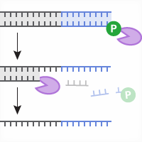
Figure 1. Engineering CRISPR-mediated gene knockins via homology-directed repair. Following formation of a double-stranded break at a genomic target site via Cas9-gRNA-mediated cleavage, a DNA template sharing homology with the targeted region can trigger homology-directed repair (HDR), resulting in knockin of exogenous sequences encoded in the DNA template (purple).

This approach has been used to engineer small edits as short as single-nucleotide substitutions, and also for the insertion of longer sequences, including fluorescent reporters or gene promoters. Knockin efficiencies can vary widely, depending in part on the species or cell type, the genomic region being targeted, and the size of the insertion. A common method for delivery of the reagents involves coelectroporation of Cas9-gRNA ribonucleoprotein complexes (RNPs) in tandem with DNA HDR templates, because it can be applied to a wide array of cell types and the transience of Cas9-gRNA expression reduces the likelihood of off-target effects. HDR templates consisting of either dsDNA or ssDNA can be used, although ssDNA has been associated with lower toxicity and a reduced likelihood of off-target integration relative to dsDNA (Roth et al., 2018). Smaller ssDNA templates (i.e., ≤200 nt) are typically obtained via oligo synthesis. For longer ssDNA, however, synthesis can be cost prohibitive, and an increasingly popular method is the Guide-it Long ssDNA Production System, which allows for on-demand production of high-quality ssDNA up to 5 kb in length from a double-stranded PCR product via selective digestion of either the sense or antisense strand. 

A powerful application of this CRISPR-mediated knockin technology involves site-specific insertion of fluorescent protein sequences to obtain fusion proteins that are expressed under the same promoter and subject to the same regulatory and cell-signaling mechanisms as their endogenous counterparts, as opposed to expressing the fusion protein from a plasmid under a different promoter (e.g., CMV) or from a non-native genomic context via random integration. To demonstrate the suitability of the Guide-it Long ssDNA Production System for this application, we used it to generate two independent hiPS cell lines expressing AcGFP1 fused to the N-termini of SEC61B and Tubulin, respectively.

Results  

Design and production of ssDNA templates

Sense and antisense single-stranded DNA (ssDNA) templates for AcGFP1 knockin at the SEC61B and TUBA1A loci were designed to include homology arms of ~350 nt flanking the AcGFP1 sequence (Figure 2), and were produced using the Guide-it Long ssDNA Production System. For comparison purposes, unprocessed dsDNA PCR products corresponding to each HDR template were retained for electroporation in parallel. It has been demonstrated that there is an exponential relationship between homology arm length and knockin efficiency, with ssDNA templates including homology arms 350–700 nt in length providing optimal performance (Li et al., 2018). In our own studies involving hiPSCs, optimal performance was observed with 350-nt homology arms, and the use of longer arms did not substantially increase knockin efficiencies.

Design of ssDNA template for knockin of AcGFP1 at SEC61B.

Figure 2. Schematics depicting Cas9-sgRNA cleavage sites and ssDNA constructs designed for knockin of AcGFP1 at the SEC61B and TUBA1A loci. The ssDNA constructs included the AcGFP1 coding sequence (~700 nt in length) flanked on either side by ~350-nt homology arms. Binding sites for PCR primers used to detect the inserted AcGFP1 sequence at the SEC61B locus are depicted by blue arrows. As indicated, neither of the ssDNA constructs included a promoter sequence, such that expression of the resulting AcGFP1-tagged fusion proteins would be driven by the respective endogenous promoters.

Electroporation of hiPSCs with ssDNA results in markedly lower toxicity vs. dsDNA

Coelectroporation of Cas9 complexed with single guide RNA (sgRNA) in tandem with HDR templates was performed according to the protocol specified for the Neon system in the Guide-it Recombinant Cas9 (Electroporation-Ready) User Manual. Cas9-sgRNA RNPs were prepared by co-incubating recombinant Cas9 protein with in vitro-transcribed sgRNAs designed to target the SEC61B or TUBA1A locus, and the RNPs were then combined with corresponding HDR templates consisting of either dsDNA or ssDNA (sense or antisense). The various RNP-HDR template combinations and negative-control RNPs without associated HDR templates were then electroporated into ChiPSC18 cells using standard conditions. After 48 hours of culturing, imaging was performed on each cell population to assess the relative toxicity associated with each RNP-HDR template combination (Figure 3). While coelectroporation of ssDNA or dsDNA HDR templates and RNPs yielded noticeable toxicity relative to the application of RNPs alone, the level of toxicity associated with dsDNA was much greater than what was observed for ssDNA. This finding is consistent with the results of previous studies performed by Takara Bio and by others (Roth et al., 2018).

Electroporation of ssDNA results in lower toxicity relative to electroporation of dsDNA.

Figure 3. Toxicity comparison for electroporation of dsDNA or ssDNA into iPS cells. ChiPSC18 cells were electroporated with Cas9-sgRNA RNPs targeting the TUBA1A locus in the absence or presence of homology-directed repair (HDR) templates consisting of dsDNA or ssDNA, and then cultured for 48 hours. While electroporation of either DNA template resulted in increased toxicity compared to electroporation of Cas9-sgRNA RNPs alone (as evidenced by lower cell densities), application of ssDNA resulted in greatly reduced toxicity relative to dsDNA.

Analysis of AcGFP1 knockin efficiencies by flow cytometry

Following culturing, the edited populations were analyzed using flow cytometry (Figure 4). In these analyses, the percentage of fluorescent-positive cells is equivalent to the percentage of successful knockins (i.e., knockin efficiency). For each locus, it was observed that the use of sense or antisense ssDNA templates yielded different knockin efficiencies; in the case of SEC61B, the sense template resulted in a higher knockin efficiency than the antisense template (12% vs. 7%, respectively; Figure 4 and data not shown), whereas for TUBA1A, the antisense template provided a higher efficiency (2.7% vs. 1.3%; Figure 4 and data not shown). Currently, there is no clear rule regarding the preferential use of sense or antisense ssDNA for HDR templates longer than 200 nt, but in the case of shorter single-stranded oligodeoxynucleotide (ssODN) templates, template polarity has been found to have an effect on efficiency (Bollen et al., 2018).

FACS analysis of AcGFP1 knockin efficiences at SEC61B and TUBA1A.

Figure 4. Analysis by flow cytometry of AcGFP1 knockin efficiencies at SEC61B and TUBA1A in iPS cells using ssDNA donors. Following electroporation and culturing, edited cell populations and unelectroporated negative control cells were analyzed for AcGFP1 expression by flow cytometry. Measuring the proportion of fluorescent cells in each population allowed for determination of knockin efficiencies: 13% knockin efficiency at SEC61B using a sense ssDNA, and 2.75% efficiency at TUBA1A using an antisense ssDNA.   

From the AcGFP1-positive populations sorted by flow cytometry, single cells were isolated using limiting dilution and expanded into clonal cell lines using the Cellartis iPSC rCas9 Electroporation and Single-Cell Cloning System.

Detection of monoallelic or biallelic knockin by PCR

One of the most important steps in characterizing edited clonal cell lines involves determining their zygosity. When the inserted sequence is of sufficient length (e.g., AcGFP1), zygosity can be easily assessed via PCR amplification of the target locus using primers that anneal outside of the genomic region that shares homology with the HDR template (Figure 5). In such analyses, a wild-type allele will generate a shorter amplicon than an allele carrying a successful knockin. To determine the zygosity of the different clonal cell lines encoding for an AcGFP1 fusion to SEC61B, genomic DNA was extracted from several of the cell lines and amplified by PCR. Agarose gel analysis of the resulting PCR products indicated that the majority of the clonal cell lines carried biallelic knockins and that comparable editing outcomes were obtained using sense (ss) or antisense (as) ssDNA templates (Figure 5).  

PCR analysis of AcGFP1 knockin at the SEC61B locus in clonal populations isolated by FACS.

Figure 5. Detection of monoallelic or biallelic knockin by PCR amplification of the target site. Genomic DNA was extracted and analyzed by PCR using forward and reverse primers designed to anneal outside the genomic region sharing homology with the HDR template. The AcGFP1 and homology arm sequences encoded in the ssDNA templates are shown in green and light blue, respectively. The PCR primers were designed such that presence or absence of an inserted AcGFP1 sequence would yield PCR products of different sizes (1.9 kb or 1.2 kb, respectively). Gel electrophoresis of PCR products confirmed the occurrence of monoallelic AcGFP1 knockins, represented by double bands (e.g., ssDNA-as, Lane C5), and biallelic AcGFP1 knockins, represented by single bands corresponding to the larger product size (e.g., ssDNA-as, Lane C1). Two wild-type clones lacking any copies of the AcGFP1 insert were also identified (ssDNA-as, Lane C10 and ssDNA-ss, Lane C11).

Analysis of AcGFP1-tagged protein localization by fluorescence microscopy

Following isolation and expansion, AcGFP1-positive clones were imaged using fluorescence microscopy (Figure 6). In each clonal population, AcGFP1 fluorescence matched the canonical localization pattern of the protein encoded by the intended genomic target: targeting of the SEC61B locus yielded AcGFP1 expression in endosomal compartments, while targeting of the TUBA1A locus resulted in fluorescent labelling of microtubules. These results suggest that electroporation of RNPs and ssDNA templates targeting SEC61B or TUBA1A resulted in successful insertion of AcGFP1 at the corresponding locus, such that the resulting fusion proteins localize in the same manner as their untagged, endogenous counterparts.

In vivo expression of AcGFP1 fusion proteins in cells edited using Cas9-sgRNA RNPs and ssDNA HDR templates.

Figure 6. Analysis of AcGFP1 localization in clonal cell lines following editing. AcGFP1-positive clonal cell lines derived from each edited population were fixed and counterstained with DAPI and analyzed by fluorescence microscopy. In each case, AcGFP1 localized in a manner consistent with the function of the endogenous protein: endosomal compartments (SEC61B locus) and microtubules (TUBA1A locus), respectively.

Conclusions  

The combination of CRISPR/Cas technology with the application of DNA templates for homology-directed repair has given rise to a powerful method for precise genome editing that can be employed in a wide array of experimental contexts. However, the efficiency of this approach varies greatly, depending on the cell type(s) and the genomic loci being targeted, and a persistent challenge involves delivering HDR templates in a manner that minimizes toxicity and the likelihood of off-target integration. Here we have presented data demonstrating that electroporation of Cas9-sgRNA RNPs in tandem with ssDNA generated using the Guide-it Long ssDNA Production System enables efficient and accurate knockin of long sequences in hiPSCs with minimal toxicity. Given the relative simplicity and cost-effectiveness of the approach presented here, we anticipate that it will continue to drive greater adoption and application of CRISPR/Cas9 technology for precise genome editing. 

References  

Bollen et al., How to create state-of-the-art genetic model systems: strategies for optimal CRISPR-mediated genome editing. Nuc. Acids Res. 46, 6435–6454 (2018).

Hsu et al., Development and applications of CRISPR-Cas9 for genome engineering. Cell 157, 1262–1278 (2014).

Li et al., Design and specificity of long ssDNA donors for CRISPR-based knockin. bioRxiv, https://doi.org/10.1101/178905 (2017).

Richardson et al., CRISPR-Cas9 genome editing in human cells occurs via the Fanconi anemia pathway. Nat. Gen. 50, 1132–1139 (2018).

Roth et al., Reprogramming human T cell function and specificity with non-viral genome targeting. Nat. Lett. 559, 405–409 (2018).

Related Products

Cat. # Product Size Price License Quantity Details

Takara Bio USA, Inc.
United States/Canada: +1.800.662.2566 • Asia Pacific: +1.650.919.7300 • Europe: +33.(0)1.3904.6880 • Japan: +81.(0)77.565.6999
FOR RESEARCH USE ONLY. NOT FOR USE IN DIAGNOSTIC PROCEDURES. © 2025 Takara Bio Inc. All Rights Reserved. All trademarks are the property of Takara Bio Inc. or its affiliate(s) in the U.S. and/or other countries or their respective owners. Certain trademarks may not be registered in all jurisdictions. Additional product, intellectual property, and restricted use information is available at takarabio.com.

Takara Bio

Takara Bio USA, Inc. provides kits, reagents, instruments, and services that help researchers explore questions about gene discovery, regulation, and function. As a member of the Takara Bio Group, Takara Bio USA is part of a company that holds a leadership position in the global market and is committed to improving the human condition through biotechnology. Our mission is to develop high-quality innovative tools and services to accelerate discovery.

FOR RESEARCH USE ONLY. NOT FOR USE IN DIAGNOSTIC PROCEDURES (EXCEPT AS SPECIFICALLY NOTED).

Support
  • Contact us
  • Technical support
  • Customer service
  • Shipping & delivery
  • Sales
  • Feedback
Products
  • New products
  • Special offers
  • Instrument & reagent services
Learning centers
  • NGS
  • Gene function
  • Stem cell research
  • Protein research
  • PCR
  • Cloning
  • Nucleic acid purification
About
  • Our brands
  • Careers
  • Events
  • Blog
  • Need help?
  • Announcements
  • Quality and compliance
  • That's Good Science!
Facebook Twitter  LinkedIn

logo strip white

©2025 Takara Bio Inc. All Rights Reserved.

Region - North America Privacy Policy Terms and Conditions Terms of Use

Top


  • COVID-19 research
  • Viral detection with qPCR
  • SARS-CoV-2 pseudovirus
  • Human ACE2 stable cell line
  • Viral RNA isolation
  • Viral and host sequencing
  • Vaccine development
  • CRISPR screening
  • Drug discovery
  • Immune profiling
  • Publications
  • Next-generation sequencing
  • Spatial omics
  • RNA-seq
  • DNA-seq
  • Single-cell NGS automation
  • Reproductive health
  • Bioinformatics tools
  • Immune profiling
  • Real-time PCR
  • Great value master mixes
  • Signature enzymes
  • High-throughput real-time PCR solutions
  • Detection assays
  • References, standards, and buffers
  • Stem cell research
  • Media, differentiation kits, and matrices
  • Stem cells and stem cell-derived cells
  • mRNA and cDNA synthesis
  • In vitro transcription
  • cDNA synthesis kits
  • Reverse transcriptases
  • RACE kits
  • Purified cDNA & genomic DNA
  • Purified total RNA and mRNA
  • PCR
  • Most popular polymerases
  • High-yield PCR
  • High-fidelity PCR
  • GC rich PCR
  • PCR master mixes
  • Cloning
  • In-Fusion seamless cloning
  • Competent cells
  • Ligation kits
  • Restriction enzymes
  • Nucleic acid purification
  • Automated platforms
  • Plasmid purification kits
  • Genomic DNA purification kits
  • DNA cleanup kits
  • RNA purification kits
  • Gene function
  • Gene editing
  • Viral transduction
  • Fluorescent proteins
  • T-cell transduction and culture
  • Tet-inducible expression systems
  • Transfection reagents
  • Cell biology assays
  • Protein research
  • Purification products
  • Two-hybrid and one-hybrid systems
  • Mass spectrometry reagents
  • Antibodies and ELISAs
  • Primary antibodies and ELISAs by research area
  • Fluorescent protein antibodies
  • New products
  • Special offers
  • In-Fusion Snap Assembly EcoDry promo
  • OEM
  • Portfolio
  • Process
  • Facilities
  • Request samples
  • FAQs
  • Instrument services
  • Apollo services
  • ICELL8 services
  • SmartChip ND system services
  • Gene and cell therapy manufacturing services
  • Services
  • Facilities
  • Our process
  • Resources
  • Customer service
  • Sales
  • Make an appointment with your sales rep
  • Shipping & delivery
  • Technical support
  • Feedback
  • Online tools
  • GoStix Plus FAQs
  • Partnering & Licensing
  • Vector information
  • Vector document overview
  • Vector document finder
Takara Bio's award-winning GMP-compliant manufacturing facility in Kusatsu, Shiga, Japan.

Partner with Takara Bio!

Takara Bio is proud to offer GMP-grade manufacturing capabilities at our award-winning facility in Kusatsu, Shiga, Japan.

  • Automation systems
  • Shasta Single Cell System introduction
  • SmartChip Real-Time PCR System introduction
  • ICELL8 introduction
  • Next-generation sequencing
  • RNA-seq
  • Technical notes
  • Technology and application overviews
  • FAQs and tips
  • DNA-seq protocols
  • Bioinformatics resources
  • Webinars
  • Spatial biology
  • Real-time PCR
  • Download qPCR resources
  • Overview
  • Reaction size guidelines
  • Guest webinar: extraction-free SARS-CoV-2 detection
  • Technical notes
  • Nucleic acid purification
  • Nucleic acid extraction webinars
  • Product demonstration videos
  • Product finder
  • Plasmid kit selection guide
  • RNA purification kit finder
  • mRNA and cDNA synthesis
  • mRNA synthesis
  • cDNA synthesis
  • PCR
  • Citations
  • PCR selection guide
  • Technical notes
  • FAQ
  • Cloning
  • Automated In-Fusion Cloning
  • In-Fusion Cloning general information
  • Primer design and other tools
  • In‑Fusion Cloning tips and FAQs
  • Applications and technical notes
  • Stem cell research
  • Overview
  • Protocols
  • Technical notes
  • Gene function
  • Gene editing
  • Viral transduction
  • T-cell transduction and culture
  • Inducible systems
  • Cell biology assays
  • Protein research
  • Capturem technology
  • Antibody immunoprecipitation
  • His-tag purification
  • Other tag purification
  • Expression systems
  • Antibodies and ELISA
  • Molecular diagnostics
  • Interview: adapting to change with Takara Bio
  • Applications
  • Solutions
  • Partnering
  • Contact us
  • mRNA and protein therapeutics
  • Characterizing the viral genome and host response
  • Identifying and cloning protein targets
  • Expressing and purifying protein targets
  • Immunizing mice and optimizing vaccines
  • Pathogen detection
  • Sample prep
  • Detection methods
  • Identification and characterization
  • SARS-CoV-2
  • Antibiotic-resistant bacteria
  • Food crop pathogens
  • Waterborne disease outbreaks
  • Viral-induced cancer
  • Immunotherapy research
  • T-cell therapy
  • Antibody therapeutics
  • T-cell receptor profiling
  • TBI initiatives in cancer therapy
  • Cancer research
  • Kickstart your cancer research with long-read sequencing
  • Sample prep from FFPE tissue
  • Sample prep from plasma
  • Cancer biomarker quantification
  • Single cancer cell analysis
  • Cancer transcriptome analysis
  • Cancer genomics and epigenomics
  • HLA typing in cancer
  • Gene editing for cancer therapy/drug discovery
  • Alzheimer's disease research
  • Antibody engineering
  • Sample prep from FFPE tissue
  • Single-cell sequencing
  • Reproductive health technologies
  • Embgenix FAQs
  • Preimplantation genetic testing
  • ESM partnership program
  • ESM Collection Kit forms
  • Infectious diseases
  • Develop vaccines for HIV
Create a web account with us

Log in to enjoy additional benefits

Want to save this information?

An account with takarabio.com entitles you to extra features such as:

•  Creating and saving shopping carts
•  Keeping a list of your products of interest
•  Saving all of your favorite pages on the site*
•  Accessing restricted content

*Save favorites by clicking the star () in the top right corner of each page while you're logged in.

Create an account to get started

  • BioView blog
  • Automation
  • Cancer research
  • Career spotlights
  • Current events
  • Customer stories
  • Gene editing
  • Research news
  • Single-cell analysis
  • Stem cell research
  • Tips and troubleshooting
  • Women in STEM
  • That's Good Support!
  • About our blog
  • That's Good Science!
  • SMART-Seq Pro Biomarker Discovery Contest
  • DNA extraction educational activity
  • That's Good Science Podcast
  • Season one
  • Season two
  • Season three
  • Our brands
  • Our history
  • In the news
  • Events
  • Biomarker discovery events
  • Calendar
  • Conferences
  • Speak with us
  • Careers
  • Company benefits
  • Trademarks
  • License statements
  • Quality statement
  • HQ-grade reagents
  • International Contacts by Region
  • United States and Canada
  • China
  • Japan
  • Korea
  • Europe
  • India
  • Affiliates & distributors
  • Need help?
  • Privacy request
  • Website FAQs

That's GOOD Science!

What does it take to generate good science? Careful planning, dedicated researchers, and the right tools. At Takara Bio, we thoughtfully develop exceptional products to tackle your most challenging research problems, and have an expert team of technical support professionals to help you along the way, all at superior value.

Explore what makes good science possible

 Customer Login
 View Cart (0)
Takara Bio
  • Home
  • Products
  • Services & Support
  • Learning centers
  • APPLICATIONS
  • About
  • Contact Us
  •  Customer Login
  • Register
  •  View Cart (0)

Takara Bio USA, Inc. provides kits, reagents, instruments, and services that help researchers explore questions about gene discovery, regulation, and function. As a member of the Takara Bio Group, Takara Bio USA is part of a company that holds a leadership position in the global market and is committed to improving the human condition through biotechnology. Our mission is to develop high-quality innovative tools and services to accelerate discovery.

FOR RESEARCH USE ONLY. NOT FOR USE IN DIAGNOSTIC PROCEDURES (EXCEPT AS SPECIFICALLY NOTED).

Clontech, TaKaRa, cellartis

  • Products
  • COVID-19 research
  • Next-generation sequencing
  • Real-time PCR
  • Stem cell research
  • mRNA and cDNA synthesis
  • PCR
  • Cloning
  • Nucleic acid purification
  • Gene function
  • Protein research
  • Antibodies and ELISA
  • New products
  • Special offers
  • COVID-19 research
  • Viral detection with qPCR
  • SARS-CoV-2 pseudovirus
  • Human ACE2 stable cell line
  • Viral RNA isolation
  • Viral and host sequencing
  • Vaccine development
  • CRISPR screening
  • Drug discovery
  • Immune profiling
  • Publications
  • Next-generation sequencing
  • Spatial omics
  • RNA-seq
  • DNA-seq
  • Single-cell NGS automation
  • Reproductive health
  • Bioinformatics tools
  • Immune profiling
  • Real-time PCR
  • Great value master mixes
  • Signature enzymes
  • High-throughput real-time PCR solutions
  • Detection assays
  • References, standards, and buffers
  • Stem cell research
  • Media, differentiation kits, and matrices
  • Stem cells and stem cell-derived cells
  • mRNA and cDNA synthesis
  • In vitro transcription
  • cDNA synthesis kits
  • Reverse transcriptases
  • RACE kits
  • Purified cDNA & genomic DNA
  • Purified total RNA and mRNA
  • PCR
  • Most popular polymerases
  • High-yield PCR
  • High-fidelity PCR
  • GC rich PCR
  • PCR master mixes
  • Cloning
  • In-Fusion seamless cloning
  • Competent cells
  • Ligation kits
  • Restriction enzymes
  • Nucleic acid purification
  • Automated platforms
  • Plasmid purification kits
  • Genomic DNA purification kits
  • DNA cleanup kits
  • RNA purification kits
  • Gene function
  • Gene editing
  • Viral transduction
  • Fluorescent proteins
  • T-cell transduction and culture
  • Tet-inducible expression systems
  • Transfection reagents
  • Cell biology assays
  • Protein research
  • Purification products
  • Two-hybrid and one-hybrid systems
  • Mass spectrometry reagents
  • Antibodies and ELISA
  • Primary antibodies and ELISAs by research area
  • Fluorescent protein antibodies
  • Special offers
  • In-Fusion Snap Assembly EcoDry promo
  • Services & Support
  • OEM
  • Instrument services
  • Gene and cell therapy manufacturing
  • Customer service
  • Sales
  • Shipping & delivery
  • Technical support
  • Feedback
  • Online tools
  • Partnering & Licensing
  • Vector information
  • OEM
  • Portfolio
  • Process
  • Facilities
  • Request samples
  • FAQs
  • Instrument services
  • Apollo services
  • ICELL8 services
  • SmartChip ND system services
  • Gene and cell therapy manufacturing
  • Services
  • Facilities
  • Our process
  • Resources
  • Sales
  • Make an appointment with your sales rep
  • Online tools
  • GoStix Plus FAQs
  • Vector information
  • Vector document overview
  • Vector document finder
  • Learning centers
  • Automation systems
  • Next-generation sequencing
  • Spatial biology
  • Real-time PCR
  • Nucleic acid purification
  • mRNA and cDNA synthesis
  • PCR
  • Cloning
  • Stem cell research
  • Gene function
  • Protein research
  • Antibodies and ELISA
  • Automation systems
  • Shasta Single Cell System introduction
  • SmartChip Real-Time PCR System introduction
  • ICELL8 introduction
  • Next-generation sequencing
  • RNA-seq
  • Technical notes
  • Technology and application overviews
  • FAQs and tips
  • DNA-seq protocols
  • Bioinformatics resources
  • Webinars
  • Real-time PCR
  • Download qPCR resources
  • Overview
  • Reaction size guidelines
  • Guest webinar: extraction-free SARS-CoV-2 detection
  • Technical notes
  • Nucleic acid purification
  • Nucleic acid extraction webinars
  • Product demonstration videos
  • Product finder
  • Plasmid kit selection guide
  • RNA purification kit finder
  • mRNA and cDNA synthesis
  • mRNA synthesis
  • cDNA synthesis
  • PCR
  • Citations
  • PCR selection guide
  • Technical notes
  • FAQ
  • Cloning
  • Automated In-Fusion Cloning
  • In-Fusion Cloning general information
  • Primer design and other tools
  • In‑Fusion Cloning tips and FAQs
  • Applications and technical notes
  • Stem cell research
  • Overview
  • Protocols
  • Technical notes
  • Gene function
  • Gene editing
  • Viral transduction
  • T-cell transduction and culture
  • Inducible systems
  • Cell biology assays
  • Protein research
  • Capturem technology
  • Antibody immunoprecipitation
  • His-tag purification
  • Other tag purification
  • Expression systems
  • APPLICATIONS
  • Molecular diagnostics
  • mRNA and protein therapeutics
  • Pathogen detection
  • Immunotherapy research
  • Cancer research
  • Alzheimer's disease research
  • Reproductive health technologies
  • Infectious diseases
  • Molecular diagnostics
  • Interview: adapting to change with Takara Bio
  • Applications
  • Solutions
  • Partnering
  • Contact us
  • mRNA and protein therapeutics
  • Characterizing the viral genome and host response
  • Identifying and cloning protein targets
  • Expressing and purifying protein targets
  • Immunizing mice and optimizing vaccines
  • Pathogen detection
  • Sample prep
  • Detection methods
  • Identification and characterization
  • SARS-CoV-2
  • Antibiotic-resistant bacteria
  • Food crop pathogens
  • Waterborne disease outbreaks
  • Viral-induced cancer
  • Immunotherapy research
  • T-cell therapy
  • Antibody therapeutics
  • T-cell receptor profiling
  • TBI initiatives in cancer therapy
  • Cancer research
  • Kickstart your cancer research with long-read sequencing
  • Sample prep from FFPE tissue
  • Sample prep from plasma
  • Cancer biomarker quantification
  • Single cancer cell analysis
  • Cancer transcriptome analysis
  • Cancer genomics and epigenomics
  • HLA typing in cancer
  • Gene editing for cancer therapy/drug discovery
  • Alzheimer's disease research
  • Antibody engineering
  • Sample prep from FFPE tissue
  • Single-cell sequencing
  • Reproductive health technologies
  • Embgenix FAQs
  • Preimplantation genetic testing
  • ESM partnership program
  • ESM Collection Kit forms
  • Infectious diseases
  • Develop vaccines for HIV
  • About
  • BioView blog
  • That's Good Science!
  • Our brands
  • Our history
  • In the news
  • Events
  • Careers
  • Trademarks
  • License statements
  • Quality and compliance
  • HQ-grade reagents
  • International Contacts by Region
  • Need help?
  • Website FAQs
  • BioView blog
  • Automation
  • Cancer research
  • Career spotlights
  • Current events
  • Customer stories
  • Gene editing
  • Research news
  • Single-cell analysis
  • Stem cell research
  • Tips and troubleshooting
  • Women in STEM
  • That's Good Support!
  • About our blog
  • That's Good Science!
  • SMART-Seq Pro Biomarker Discovery Contest
  • DNA extraction educational activity
  • That's Good Science Podcast
  • Season one
  • Season two
  • Season three
  • Events
  • Biomarker discovery events
  • Calendar
  • Conferences
  • Speak with us
  • Careers
  • Company benefits
  • International Contacts by Region
  • United States and Canada
  • China
  • Japan
  • Korea
  • Europe
  • India
  • Affiliates & distributors
  • Need help?
  • Privacy request
Takara Bio
  • Products
  • Services & Support
  • Learning centers
  • APPLICATIONS
  • About
  • Contact Us