We use cookies to improve your browsing experience and provide meaningful content. Read our cookie policy. Accept
  •  Customer Login
  • Register
  •  View Cart (0)
  •  Customer Login
  • Register
  •  View Cart (0)

  • Products
  • Services & Support
  • Learning centers
  • APPLICATIONS
  • About
  • Contact Us

Close

  • ‹ Back to Creating and screening for SNPs
  • SNP detection with knockin screening kit
  • Oligo design tool for SNP screening
  • Sign up: SNP engineering webinar
  • Guide-it SNP Screening Kit FAQs
Guide-it Knockin Screening Kit product page Guide-it Knockin Screening Kit product information
Oligo design tool for assaying SNPs Oligo design tool for assaying SNPs
Home › Learning centers › Gene function › Gene editing › Creating and screening for SNPs › SNP detection with knockin screening kit

Gene editing

  • Gene editing product finder
  • Gene editing tools and information
    • sgRNA design tools
    • Tools for successful CRISPR/Cas9 genome editing
    • Gene editing posters
    • Customer data for Guide-it products
    • How to design sgRNA sequences
    • Introduction to the CRISPR/Cas9 system
    • Gene editing of CD3+ T cells and CD34+ HSCs
  • CRISPR/Cas9 knockouts
    • Mutation detection kit comparison
    • Screening for effective guide RNAs
    • Monoallelic versus biallelic mutants
    • Indel identification kit for mutation characterization
  • CRISPR/Cas9 knockins
    • Choosing an HDR template format
    • Homology-directed repair FAQs
    • Mouse CRISPR knockin protocol
    • Site-specific gene knockins using long ssDNA
    • Efficient CRISPR/Cas9-mediated knockins in iPS cells
    • Oligo design tool for detecting precise insertions
      • Oligo design tool user guide (insertions)
  • Genome-wide screening
    • CRISPR library screening
    • CRISPR library screening webinar
    • Phenotypic screen using sgRNA library system
  • Creating and screening for SNPs
    • SNP detection with knockin screening kit
    • Oligo design tool for SNP screening
      • Oligo design tool user guide (SNPs)
    • Sign up: SNP engineering webinar
    • Guide-it SNP Screening Kit FAQs
  • CRISPR/Cas9 delivery methods
    • Electroporation-grade Cas9 for editing in diverse cell types
    • CRISPR/Cas9 gene editing with AAV
    • CRISPR/Cas9 gesicles overview
    • Cas9 Gesicles—reduced off-target effects
    • sgRNA-Cas9 delivery to many cell types
    • Tet-inducible Cas9 for gene editing
  • Cre recombinase
    • Control your Cre recombinase experiments
    • Fast Cre delivery with gesicle technology
New products
Need help?
Contact Sales
Guide-it Knockin Screening Kit product page Guide-it Knockin Screening Kit product information
Oligo design tool for assaying SNPs Oligo design tool for assaying SNPs
Tech Note

SNP engineering with the Guide-it Knockin Screening Kit

  • Genome editing technologies enable the generation of disease models by combining site-specific DNA cleavage with homology-directed repair (HDR) to engineer disease-associated SNPs
  • The efficiency of accurate HDR can vary widely, and the identification of successfully edited clones using existing methods can prove challenging
  • The Guide-it Knockin Screening Kit provides a fast and accurate assay for detecting the successful introduction of SNPs, both in mixed populations and in clonal cell lines
  • Convenient online tool available for easy, error-free design of assay-specific oligo probes
  • The dual-color technology employed by the kit allows simultaneous detection of two alleles, enabling identification of heterozygous cell lines carrying one copy each of SNP and WT alleles
  • The kit can be used to screen for clones carrying the desired edit and yields more accurate and easier-to-interpret results than bioinformatic analysis of Sanger sequencing data

Introduction Workflow In-house data Beta-tester data Conclusions

Introduction  

The ease with which somatic cells obtained from patient and nonpatient populations can be reprogrammed into human induced pluripotent stem cells (iPSCs) and edited in a precise manner (using CRISPR or related technologies) has dramatically expanded the applicability and accessibility of iPSC-derived cell lines for disease modeling and drug development (Figure 1). Introduction of disease-associated genetic variants, including SNPs, in iPSCs allows for the evaluation of the phenotypic effects of these variants in relevant cell types and eliminates variability due to genetic background, thus enabling finer analysis of disease mechanisms and more rapid assessment of potential therapies (e.g., drug screening). Looking a step further, a promising clinical application for precise genome editing could be the correction of pathogenic mutations in patient-derived iPSCs and delivery of the engineered cells to the patients as therapy.

Applications for genome editing in iPS cells

Figure 1. Applications for genome editing in iPSCs. The genetic modification of iPSC lines to introduce pathogenic mutations or correct them has enabled the generation of isogenic cell lines for disease modeling and drug screening applications. In the future, the correction of disease-associated mutations in patient-derived iPSCs could also provide a basis for novel therapeutic approaches. Image from Kim, Kang & Ju, 2017 used under creative commons license. Copyright @ 2017 The Korean Association of Internal Medicine.

Workflow  

One of the most common strategies in creating disease models is the use of CRISPR/Cas9 in tandem with single-stranded donor oligonucleotide (ssODN) encoding the desired modification, but this approach relies on homology-directed repair (HDR), an inefficient endogenous repair mechanism that typically results in a low percentage of successfully edited cells.

Genome editing experiments involving the application of HDR to generate SNPs typically yield three possible outcomes at the target site: retention of the wild-type (WT) sequence (i.e., no editing), creation of an insertion or deletion (Indel), or successful HDR (SNP) (Figure 2, Panel A). These outcomes can be distributed in varying allelic combinations in diploid cells (Figure 2, Panel B). When studying diseases caused by dominant alleles, researchers often want to generate isogenic cell lines with one allele edited (encoding the SNP of interest) while maintaining the other allele as wild type. This outcome is particularly difficult to achieve since most cells edited via HDR are either homozygous for the SNP (SNP/SNP) or carry an indel allele in addition to the SNP allele (SNP/Indel). This challenge, together with the low efficiency of HDR, has necessitated the optimization of the editing conditions (to maximize HDR efficiency) and the development of fast and reliable methods for screening large numbers of clonal cell lines.

Common outcomes when engineering SNPs.

Figure 2. Common outcomes when engineering SNPs. An example of a single-nucleotide edit (G>T) is shown. Panel A. Outcomes at the genomic target site. When cleavage fails to occur at the target site or is followed by accurate, nonhomologous end joining (NHEJ)-based repair, the result is the wild-type (WT) sequence. When cleavage is followed by inaccurate NHEJ-based repair, the result is an insertion or deletion (Indel) at the target site, possibly causing a knockout (KO; a highly probable outcome). When cleavage is followed by accurate HDR, a SNP is introduced at the target site. Panel B. Combined allelic outcomes in diploid cells. When editing is performed in diploid cells, the outcomes for each allele can vary, generating multiple possible combinations. Cells can remain homozygous (Wild type; top), they can have one or both alleles modified via inaccurate NHEJ (Indel; middle), or they can have one or both alleles modified with the desired SNP (Successful HDR; bottom).

The Guide-it Knockin Screening Kit offers the possibility to directly analyze HDR efficiency in bulk-edited populations, allowing researchers to move forward with isolation of single-cell clones from populations that have the highest frequency of successful HDR. Subsequently, clonal cell lines can be screened simultaneously for two specific edits (e.g., SNP/WT).

The first step of the workflow for the Guide-it Knockin Screening Kit involves PCR amplification of the targeted genomic locus. The resulting PCR product is then hybridized with two user-designed oligos encoding the sequence to be detected. If there is full hybridization between the oligos and the PCR product, an enzymatic reaction that generates a fluorescent signal is triggered, such that detection of a fluorescent signal is correlated with the presence of a specific sequence at the target site. The assay is sensitive enough to detect single-base differences, and the dual fluorescence capability (red/green) of the kit allows for the simultaneous detection of two different sequences at the same target site (Figure 3). Therefore, the kit can be used to screen for heterozygous clones with one probe encoding the SNP allele (triggering a green signal) and another probe encoding the wild-type allele (generating a red signal).

SNP analysis workflow for the Guide-it Knockin Screening Kit.

Figure 3. SNP analysis workflow for the Guide-it Knockin Screening Kit. This example workflow demonstrates analysis of a G>A substitution, where G is the wild-type base edited to an A. After genome editing, single cells expanded to clonal cell lines can have several different genotypic outcomes at the genomic target site of interest (as shown in Figure 2, Panel B). After PCR amplification of the target site, the PCR product is annealed simultaneously with different oligo probes: a displacement oligo (purple) in combination with either flap-probe oligo A (green; encoding the SNP allele, A) or flap-probe oligo B (orange; encoding the WT allele, G). After the annealing of the oligos to the PCR products, the Guide-it Flapase enzyme (indicated with scissors) recognizes a complete base pairing and cleaves the 5′ portion of the flap-probe oligo (shaded green or orange). The cleaved flaps are then detected by corresponding Guide-it flap detectors, which yield green or red fluorescent signals, respectively. In the example above, analysis of a clonal cell line that is homozygous WT (G/G) at the site of interest yields only a red signal, while analysis of a heterozygous clone carrying both edited and WT alleles (G/A) yields both red and green signals.

In-house data  

Detection of precise editing at a specific genomic locus in bulk-edited and clonal cell populations

One of the strategies to maximize the percentage of heterozygous clones (WT/SNP) is to use an equimolar mixture of ssODNs encoding the SNP or WT alleles and silent mutations in the PAM site (if possible) as HDR templates (Paquet et al. 2016). Mutation of the PAM site prevents Cas9 from recutting and generating indels at the genomic target site following successful HDR with either template, indirectly increasing the likelihood of obtaining heterozygotes.

We applied this method to generate an iPSC line heterozygous for a C>G substitution (S38C) in an endogenous gene of interest (unidentified for reasons of confidentiality). We electroporated two HDR templates (Figure 4, Panel A) encoding either the SNP (in blue) or the WT allele (in purple). The introduction of the SNP results in the destruction of the PAM site (CCG>GCG), and the oligo encoding the WT allele also codes for a silent mutation in the PAM sequence (C>G, in red; CCG>CGG).

The Guide-it Knockin Screening Kit was used to detect successful HDR of both templates (SNP or WT silent) in bulk-edited populations (Figure 4, Panel B). The lack of green and red fluorescent signals for cells electroporated with Cas9 protein in the absence of sgRNA ("negative control") or HDR templates ("KO") confirmed the specificity of the flap-probe oligos since they did not yield false-positive fluorescent signals in the presence of the unedited wild-type sequence or indel alleles.

Following editing and clonal cell isolation and expansion, the Guide-it Knockin Screening Kit was also used to identify clones with successful editing. Clones carrying the SNP or WT silent alleles could be detected with green or red fluorescence, respectively, but no heterozygotes carrying both edits (generating both red and green fluorescence signals; Figure 4, Panel C) were identified in this experiment, which had a low percentage of HDR.

Detection of precise editing at the TIMP3 locus in bulk-edited and clonal iPS cell populations

Figure 4. Detection of precise editing at an endogenous locus in bulk-edited and clonal iPSC populations. Panel A. Editing outcomes following successful HDR at an anonymous locus of interest. Following successful HDR, the edited locus will encode either a SNP (in blue, lowercase) or a WT base (in purple) combined with a silent PAM mutation (in red, lowercase). Panel B. Detection of successful HDR in bulk-edited iPSCs. Displacement and flap-probe oligos were designed to detect WT silent or SNP alleles, yielding red and green fluorescent signals, respectively. In independent experiments, cells were electroporated with Cas9 protein alone (negative control), Cas9-sgRNA RNP complexes (KO), or RNP complexes combined with antisense SNP or SNP/WT silent ssODN mixtures. Synthetic oligos encoding the WT silent or SNP sequences were assayed in parallel as positive controls. For each editing scenario in which ssODNs were included in the electroporation mixture, successful HDR could be detected in the bulk population using the Guide-it Knockin Screening Kit, as indicated by the resulting fluorescent signals. Panel C. Detection of successful HDR in clonal cell lines. Clones obtained from single cells isolated by flow cytometry were screened for both edits (SNP and WT silent). While successful incorporation of either edit could be detected in separate clonal cell lines, no heterozygous clones carrying both edits were identified.

Comparing the results of the knockin screening kit with direct Sanger sequencing

The results of the Guide-it knockin screening were confirmed by Sanger sequencing. In cases where direct Sanger sequencing of the PCR reaction yielded ambiguous results or traces that were challenging to interpret (e.g., for instances where one of the edited alleles contained an indel), the genomic target region was amplified by PCR and cloned into pUC19 following the protocol of the Guide-it Indel Identification Kit. Sequencing of several bacterial clones in each case confirmed the results from the Guide-it Knockin Screening Kit assay.

Comparing the results of the knockin screening kit with direct Sanger sequencing demonstrated the challenges associated with interpreting data from the latter approach, even when the Sanger data was analyzed with the aid of a commonly used bioinformatics tool. To demonstrate this, we compared results obtained using either the knockin screening kit or Sanger sequencing to analyze a clonal cell line edited at an unidentified locus as described above (Figure 5). The knockin screening kit detected the presence of at least one correctly edited WT allele (WT silent), indicating that there could be two copies of the WT silent allele or that it could be paired with an allele encoding the unedited wild-type sequence or an indel. In contrast, direct Sanger sequencing of the PCR product amplified from the clonal cell line yielded a sequencing chromatogram with overlapping peaks at the region of interest (Figure 5, first chromatogram). Analysis of this sequencing data with a commonly used bioinformatic tool indicated that different alleles could be present in the sample at various probabilities (WT, 44%; WT silent, 17%; 1-bp deletion, 14%; another 1-bp deletion, 11%). Using the Guide-it Indel Identification Kit, it was determined that the clone had one correctly edited WT allele (WT silent; Figure 5, middle chromatogram) and one indel allele with a one-base insertion (G) together with a 7-bp deletion (Figure 5, bottom chromatogram). This result was consistent with the output of the knockin screening kit and demonstrated that the results obtained via bioinformatic analysis of Sanger sequencing could be misleading.

Comparing results from the Guide-it Knockin Screening Kit vs. Sanger sequencing

Figure 5. Comparing results from the Guide-it Knockin Screening Kit vs. Sanger sequencing. The knockin screening assay detected the presence of at least one correctly edited WT allele (WT silent; top sequence). In contrast, direct Sanger sequencing of the PCR product amplified from the clonal population yielded a sequencing chromatogram with overlapping peaks at the region of interest (top chromatogram). Analysis of this sequencing data with a commonly used bioinformatics tool indicated that different alleles could be present in the sample at various probabilities. Using the Guide-it Indel Identification Kit, it was determined that the clone had one correctly edited WT allele (WT silent; middle chromatogram) and one indel allele with a one-base insertion (G) together with a 7-bp deletion (bottom chromatogram), a result consistent with the output of the Guide-it Knockin Screening Kit.

Beta-tester data  

Comparing results of the knockin screening kit with Sanger sequencing in clonal cell lines

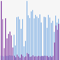
Dr. Justin McDonough, from the Jackson Laboratory, compared the performance of the Guide-it Knockin Screening Kit vs. software-based analysis of Sanger sequencing data obtained from PCR products for the detection of heterozygous (SNP/WT) edited iPSC clones (Figure 6). When interpreting these results, it must be taken into account that the detection of only one fluorescent signal (green or red) in a clonal cell line is correlated to the presence of at least one copy of the corresponding allele and indicates the absence of the other allele being assayed. For example, the genotype of a clone that generates only a green signal is SNP/SNP or SNP/Indel but not WT/SNP, WT/WT, or WT/Indel.

While the results of the Guide-it Knockin Screening Kit were consistent with the analysis of the Sanger sequencing traces for a majority of the clones (e.g., heterozygous clone A07), there were several instances where analysis of the Sanger data with a commonly used bioinformatics tool missed or called some clones incorrectly. For example, one clone carrying the SNP could not be identified by analysis of the Sanger data (F04; designated "unknown" in Figure 6). In addition, another clone carrying the desired SNP (E07) was called incorrectly to encode two copies of indel alleles at the target site. The beta tester indicated that, "the kit was very good at identifying WT/SNV hets, which is often what I'm looking to uncover."

Beta-tester data: successful identification of heterozygous edited clones.

Figure 6. Beta-tester data: successful identification of heterozygous edited clones. Genotypes determined via bioinformatic analysis of the Sanger sequencing data are indicated along the X-axis (WT, wild-type; SNP, accurate HDR; Indel, NHEJ; unknown, software could not determine). The presence of edited (SNP) and wild-type (WT) alleles in the clones as determined by the Guide-it Knockin Screening Kit are demonstrated by fluorescence signal intensities indicated in blue (green fluorescence) and purple (red fluorescence), respectively. While the results of the knockin screening were consistent with the bioinformatic analysis of the Sanger sequencing traces for a majority of clones analyzed, there were several instances where the Sanger sequencing analysis missed or called some clones incorrectly.

Conclusions  

For researchers seeking to elucidate the genetic basis of diseases or develop novel therapies, the ability to engineer disease-associated SNPs using CRISPR-based genome editing technology in tandem with HDR has opened many doors. However, this approach is not without its challenges, due in part to the tendency towards NHEJ-based repair over HDR, and the difficulty associated with identifying successful HDR via Sanger sequencing of PCR products amplified from edited cells (even with the assistance of bioinformatic software tools). The Guide-it Knockin Screening Kit was developed with these challenges in mind. It provides researchers with a simple, rapid method to unequivocally identify successful HDR, both in bulk-edited populations prior to the tedious, time-consuming isolation of single-cell clones, and in the resulting clonal populations. As demonstrated by the data presented above, the knockin screening assay identifies successful HDR with greater certainty than direct Sanger sequencing and enables simultaneous detection of two different alleles (e.g., SNP and WT), a key benefit for researchers seeking to engineer cell lines that are heterozygous for a SNP allele of interest.

References

Kim, E. J., Kang, K. H. & Ju, J. H. CRISPR-Cas9: a promising tool for gene editing on induced pluripotent stem cells. Korean J. Intern. Med. 32, 42–61 (2017).

Paquet, D. et al. Efficient introduction of specific homozygous and heterozygous mutations using CRISPR/Cas9. Nature 533, 125–129 (2016).

Related Products

Cat. # Product Size License Quantity Details
632659 Guide-it™ Knockin Screening Kit 100 Rxns USD $434.00

License Statement

ID Number  
325 Patent pending. For further information, please contact a Takara Bio USA licensing representative by email at licensing@takarabio.com.

The Guide-it Knockin Screening Kit enables sensitive detection of successful homologous recombination (HR) events in mixed or clonal cell populations edited using technologies such as the CRISPR/Cas9 system. The kit employs a simple fluorescence-based method that can reliably detect successful HR events regardless of the knockin length (from single-nucleotide substitutions to longer insertions) or the sequence of the genomic region surrounding the edit. The simple and rapid kit workflow consists of PCR amplification of the genomic target site followed by an enzymatic assay with green and red fluorescent readouts. The enzymatic assay employs a standard fluorescence plate reader or qPCR machine for endpoint detection of fluorescence, and no additional special instrumentation is required. The overall workflow takes approximately four hours to complete, and the stringency of the assay is such that detection of fluorescent signal(s) positively correlates with the presence of the desired sequence at the genomic target site. For research applications that involve engineering SNPs, the assay can be used to positively identify heterozygous clones carrying one copy each of two different alleles (e.g., SNP and WT alleles). For scenarios involving knockin of longer sequences, the assay allows for the simultaneous detection of seamless insertions at both 5' and 3' ends of the incorporated sequence.

Cat. # 632659 includes sufficient quantities of reagents for performing 100 assays.

Notice to purchaser

Our products are to be used for Research Use Only. They may not be used for any other purpose, including, but not limited to, use in humans, therapeutic or diagnostic use, or commercial use of any kind. Our products may not be transferred to third parties, resold, modified for resale, or used to manufacture commercial products or to provide a service to third parties without our prior written approval.

Documents Components Image Data

Back

Simultaneous detection of WT and SNP alleles carrying a silent PAM mutation at the PSEN1 locus in a bulk-edited iPS cell population

Simultaneous detection of WT and SNP alleles carrying a silent PAM mutation at the PSEN1 locus in a bulk-edited iPS cell population

To demonstrate the SNP-detection capabilities of the Guide-it Knockin Screening Kit, we used CRISPR/Cas editing technology to generate an iPS cell line heterozygous for a variant of the PSEN1 gene encoding an A>G substitution (M164V) associated with early-onset Alzheimer's disease. Panel A. HDR templates carrying silent PAM mutations used to perform precise editing at the PSEN1 locus. Following successful HDR, the PSEN1 locus will encode either a WT or SNP allele combined with a silent mutation in the neighboring PAM sequence. Panel B. Design of displacement and flap-probe oligos for detection of edits involving silent mutation of the PAM sequence (G>C) and introduction of the SNP (A>G). The displacement oligo probe (in purple) is designed to hybridize in a similar manner when either allele is present (HDR wt silent or HDR M164V). The two different flap-probe oligos (in orange and green) are designed to fully hybridize to either the HDR wt silent allele encoding the PAM mutation (generating a red signal) or the HDR M164V allele encoding both the PAM mutation and the SNP (generating a green signal), respectively. The fixed sequences responsible for generating the fluorescent signals are underlined for each flap-probe oligo. Assayed bases for each allele are indicated in lowercase font.

Back

Common outcomes when engineering SNPs

Common outcomes when engineering SNPs

An example of a single-nucleotide edit (G>T) is shown. Panel A. Outcomes at the genomic target site. When cleavage fails to occur at the target site or is followed by accurate, nonhomologous end joining (NHEJ)-based repair, the result is the wild-type (WT) sequence. When cleavage is followed by inaccurate NHEJ-based repair, the result is an insertion or deletion (Indel) at the target site possibly causing a knockout (KO, a highly probable outcome). When cleavage is followed by accurate HDR, a SNP is introduced at the target site. Panel B. Combined allelic outcomes in diploid cells. When editing is performed in diploid cells, the outcomes for each allele can vary, generating multiple possible combinations. Cells can remain homozygous (Wild type; top), they can have one or both alleles modified via inaccurate NHEJ (Indel; middle), or they can have one or both alleles modified with the desired SNP (Successful HDR; bottom).

Back

SNP analysis workflow for the Guide-it Knockin Screening Kit

SNP analysis workflow for the Guide-it Knockin Screening Kit

This example workflow demonstrates analysis of a G>A substitution, where G is the wild-type base edited to an A. After genome editing, single cells expanded to clonal cell lines can have several different genotypic outcomes at the genomic target site of interest. After PCR amplification of the target site, the PCR product is annealed simultaneously with different oligo probes: a displacement oligo (purple) in combination with either flap-probe oligo A (green; encoding the SNP allele, A) or flap-probe oligo B (orange; encoding the WT allele, G). After the annealing of the oligos to the PCR products, the Guide-it Flapase enzyme (indicated with scissors) recognizes a complete base pairing and cleaves the 5′ portion of the flap-probe oligo (shaded green or orange). The cleaved flaps are then detected by corresponding Guide-it flap detectors, which yield green or red fluorescent signals, respectively. In the example above, analysis of a clonal cell line that is homozygous WT (G/G) at the site of interest yields only a red signal, while analysis of a heterozygous clone carrying both edited and WT alleles (G/A) yields both red and green signals.

Back

Detection of precise editing at an endogenous locus in bulk-edited and clonal iPSC populations

Detection of precise editing at an endogenous locus in bulk-edited and clonal iPSC populations

Panel A. Editing outcomes following successful HDR at an anonymous locus of interest. Following successful HDR, the edited locus will encode either a SNP (in blue, lowercase) or a WT base (in purple) combined with a silent PAM mutation (in red, lowercase). Panel B. Detection of successful HDR in bulk-edited iPSCs. Displacement and flap-probe oligos were designed to detect WT silent or SNP alleles, yielding red and green fluorescent signals, respectively. In independent experiments, cells were electroporated with Cas9 protein alone (negative control), Cas9-sgRNA RNP complexes (KO), or RNP complexes combined with antisense SNP or SNP/WT silent ssODN mixtures. Synthetic oligos encoding the WT silent or SNP sequences were assayed in parallel as positive controls. For each editing scenario in which ssODNs were included in the electroporation mixture, successful HDR could be detected in the bulk population using the Guide-it Knockin Screening Kit, as indicated by the resulting fluorescent signals. Panel C. Detection of successful HDR in clonal cell lines. Clones obtained from single cells isolated by flow cytometry were screened for both edits (SNP and WT silent). While successful incorporation of either edit could be detected in separate clonal cell lines, no heterozygous clones carrying both edits were identified.

Back

The Guide-it Knockin Screening Kit provides a method for detecting full-length knockin insertions

The Guide-it Knockin Screening Kit provides a method for detecting full-length knockin insertions

After the genome editing event, bulk-edited population or clonal cell lines isolated via FACS or limiting dilution may carry wild-type, indel, or full-length insertions. After DNA extraction from the clonal cells and subsequent PCR amplification of the target site, the PCR product is annealed with two different sets of displacement and flap probes: one that hybridizes with the 5' end of the insert (Flap-probe oligo A; green), and the other with the 3' end (Flap-probe oligo B; orange). If the full-length HR event has been successful and seamless, the full hybridization of the probes at both termini will generate both green and red fluorescent signals after the cleavage of the respective flap probes by the Guide-it Flapase. Detection of only one signal (red or green) indicates an insertion truncated on either the 5' or 3' end, respectively. The lack of fluorescence is indicative of the presence of the wild-type sequence or an indel at the target site.

Back

Beta-tester data: successful identification of heterozygous edited clones

Beta-tester data: successful identification of heterozygous edited clones

Genotypes determined via bioinformatic analysis of the Sanger sequencing data are indicated along the X-axis (WT, wild-type; SNP, accurate HDR; Indel, NHEJ; unknown, software could not determine). The presence of edited (SNP) and wild-type (WT) alleles in the clones as determined by the Guide-it Knockin Screening Kit are demonstrated by fluorescence signal intensities indicated in blue (green fluorescence) and purple (red fluorescence), respectively. While the results of the knockin screening were consistent with the bioinformatic analysis of the Sanger sequencing traces for a majority of clones analyzed, there were several instances where the Sanger sequencing analysis missed or called some clones incorrectly.

Back

632659: Guide-it Knockin Screening Kit

632659: Guide-it Knockin Screening Kit
632660 Guide-it™ Knockin Screening Kit 400 Rxns USD $1112.00

License Statement

ID Number  
325 Patent pending. For further information, please contact a Takara Bio USA licensing representative by email at licensing@takarabio.com.

The Guide-it Knockin Screening Kit enables sensitive detection of successful homologous recombination (HR) events in mixed or clonal cell populations edited using technologies such as the CRISPR/Cas9 system. The kit employs a simple fluorescence-based method that can reliably detect successful HR events regardless of the knockin length (from single-nucleotide substitutions to longer insertions) or the sequence of the genomic region surrounding the edit. The simple and rapid kit workflow consists of PCR amplification of the genomic target site followed by an enzymatic assay with green and red fluorescent readouts. The enzymatic assay employs a standard fluorescence plate reader or qPCR machine for endpoint detection of fluorescence, and no additional special instrumentation is required. The overall workflow takes approximately four hours to complete, and the stringency of the assay is such that detection of fluorescent signal(s) positively correlates with the presence of the desired sequence at the genomic target site. For research applications that involve engineering SNPs, the assay can be used to positively identify heterozygous clones carrying one copy each of two different alleles (e.g., SNP and WT alleles). For scenarios involving knockin of longer sequences, the assay allows for the simultaneous detection of seamless insertions at both 5' and 3' ends of the incorporated sequence.

Cat. # 632660 includes sufficient quantities of reagents for performing 400 assays.

Notice to purchaser

Our products are to be used for Research Use Only. They may not be used for any other purpose, including, but not limited to, use in humans, therapeutic or diagnostic use, or commercial use of any kind. Our products may not be transferred to third parties, resold, modified for resale, or used to manufacture commercial products or to provide a service to third parties without our prior written approval.

Documents Components Image Data

Back

Simultaneous detection of WT and SNP alleles carrying a silent PAM mutation at the PSEN1 locus in a bulk-edited iPS cell population

Simultaneous detection of WT and SNP alleles carrying a silent PAM mutation at the PSEN1 locus in a bulk-edited iPS cell population

To demonstrate the SNP-detection capabilities of the Guide-it Knockin Screening Kit, we used CRISPR/Cas editing technology to generate an iPS cell line heterozygous for a variant of the PSEN1 gene encoding an A>G substitution (M164V) associated with early-onset Alzheimer's disease. Panel A. HDR templates carrying silent PAM mutations used to perform precise editing at the PSEN1 locus. Following successful HDR, the PSEN1 locus will encode either a WT or SNP allele combined with a silent mutation in the neighboring PAM sequence. Panel B. Design of displacement and flap-probe oligos for detection of edits involving silent mutation of the PAM sequence (G>C) and introduction of the SNP (A>G). The displacement oligo probe (in purple) is designed to hybridize in a similar manner when either allele is present (HDR wt silent or HDR M164V). The two different flap-probe oligos (in orange and green) are designed to fully hybridize to either the HDR wt silent allele encoding the PAM mutation (generating a red signal) or the HDR M164V allele encoding both the PAM mutation and the SNP (generating a green signal), respectively. The fixed sequences responsible for generating the fluorescent signals are underlined for each flap-probe oligo. Assayed bases for each allele are indicated in lowercase font.

Back

Common outcomes when engineering SNPs

Common outcomes when engineering SNPs

An example of a single-nucleotide edit (G>T) is shown. Panel A. Outcomes at the genomic target site. When cleavage fails to occur at the target site or is followed by accurate, nonhomologous end joining (NHEJ)-based repair, the result is the wild-type (WT) sequence. When cleavage is followed by inaccurate NHEJ-based repair, the result is an insertion or deletion (Indel) at the target site possibly causing a knockout (KO, a highly probable outcome). When cleavage is followed by accurate HDR, a SNP is introduced at the target site. Panel B. Combined allelic outcomes in diploid cells. When editing is performed in diploid cells, the outcomes for each allele can vary, generating multiple possible combinations. Cells can remain homozygous (Wild type; top), they can have one or both alleles modified via inaccurate NHEJ (Indel; middle), or they can have one or both alleles modified with the desired SNP (Successful HDR; bottom).

Back

SNP analysis workflow for the Guide-it Knockin Screening Kit

SNP analysis workflow for the Guide-it Knockin Screening Kit

This example workflow demonstrates analysis of a G>A substitution, where G is the wild-type base edited to an A. After genome editing, single cells expanded to clonal cell lines can have several different genotypic outcomes at the genomic target site of interest. After PCR amplification of the target site, the PCR product is annealed simultaneously with different oligo probes: a displacement oligo (purple) in combination with either flap-probe oligo A (green; encoding the SNP allele, A) or flap-probe oligo B (orange; encoding the WT allele, G). After the annealing of the oligos to the PCR products, the Guide-it Flapase enzyme (indicated with scissors) recognizes a complete base pairing and cleaves the 5′ portion of the flap-probe oligo (shaded green or orange). The cleaved flaps are then detected by corresponding Guide-it flap detectors, which yield green or red fluorescent signals, respectively. In the example above, analysis of a clonal cell line that is homozygous WT (G/G) at the site of interest yields only a red signal, while analysis of a heterozygous clone carrying both edited and WT alleles (G/A) yields both red and green signals.

Back

Detection of precise editing at an endogenous locus in bulk-edited and clonal iPSC populations

Detection of precise editing at an endogenous locus in bulk-edited and clonal iPSC populations

Panel A. Editing outcomes following successful HDR at an anonymous locus of interest. Following successful HDR, the edited locus will encode either a SNP (in blue, lowercase) or a WT base (in purple) combined with a silent PAM mutation (in red, lowercase). Panel B. Detection of successful HDR in bulk-edited iPSCs. Displacement and flap-probe oligos were designed to detect WT silent or SNP alleles, yielding red and green fluorescent signals, respectively. In independent experiments, cells were electroporated with Cas9 protein alone (negative control), Cas9-sgRNA RNP complexes (KO), or RNP complexes combined with antisense SNP or SNP/WT silent ssODN mixtures. Synthetic oligos encoding the WT silent or SNP sequences were assayed in parallel as positive controls. For each editing scenario in which ssODNs were included in the electroporation mixture, successful HDR could be detected in the bulk population using the Guide-it Knockin Screening Kit, as indicated by the resulting fluorescent signals. Panel C. Detection of successful HDR in clonal cell lines. Clones obtained from single cells isolated by flow cytometry were screened for both edits (SNP and WT silent). While successful incorporation of either edit could be detected in separate clonal cell lines, no heterozygous clones carrying both edits were identified.

Back

The Guide-it Knockin Screening Kit provides a method for detecting full-length knockin insertions

The Guide-it Knockin Screening Kit provides a method for detecting full-length knockin insertions

After the genome editing event, bulk-edited population or clonal cell lines isolated via FACS or limiting dilution may carry wild-type, indel, or full-length insertions. After DNA extraction from the clonal cells and subsequent PCR amplification of the target site, the PCR product is annealed with two different sets of displacement and flap probes: one that hybridizes with the 5' end of the insert (Flap-probe oligo A; green), and the other with the 3' end (Flap-probe oligo B; orange). If the full-length HR event has been successful and seamless, the full hybridization of the probes at both termini will generate both green and red fluorescent signals after the cleavage of the respective flap probes by the Guide-it Flapase. Detection of only one signal (red or green) indicates an insertion truncated on either the 5' or 3' end, respectively. The lack of fluorescence is indicative of the presence of the wild-type sequence or an indel at the target site.

Back

Beta-tester data: successful identification of heterozygous edited clones

Beta-tester data: successful identification of heterozygous edited clones

Genotypes determined via bioinformatic analysis of the Sanger sequencing data are indicated along the X-axis (WT, wild-type; SNP, accurate HDR; Indel, NHEJ; unknown, software could not determine). The presence of edited (SNP) and wild-type (WT) alleles in the clones as determined by the Guide-it Knockin Screening Kit are demonstrated by fluorescence signal intensities indicated in blue (green fluorescence) and purple (red fluorescence), respectively. While the results of the knockin screening were consistent with the bioinformatic analysis of the Sanger sequencing traces for a majority of clones analyzed, there were several instances where the Sanger sequencing analysis missed or called some clones incorrectly.

Back

632660: Guide-it Knockin Screening Kit

632660: Guide-it Knockin Screening Kit
631444 Guide-it™ Indel Identification Kit 10 Rxns USD $462.00

The Guide-it Indel Identification Kit is used for characterization of insertions and deletions (indels) generated by gene editing tools, such as CRISPR/Cas9. This kit contains all of the components needed to amplify, clone, and prepare modified target sites for DNA sequence analysis. This kit uses Terra PCR Direct to amplify targets directly from crude genomic DNA extracts. The resulting pool of fragments, which may contain a variety of indels, are cloned into a prelinearized pUC19 vector using the In-Fusion cloning system. Colony PCR of individual clones using Terra PCR Direct followed by DNA sequencing allows indel characterization.

Notice to purchaser

Our products are to be used for Research Use Only. They may not be used for any other purpose, including, but not limited to, use in humans, therapeutic or diagnostic use, or commercial use of any kind. Our products may not be transferred to third parties, resold, modified for resale, or used to manufacture commercial products or to provide a service to third parties without our prior written approval.

Documents Components Image Data Resources

Back

Identification of insertions and deletions (indels) in the CD81 gene after CRISPR/Cas9 targeting

Identification of insertions and deletions (indels) in the CD81 gene after CRISPR/Cas9 targeting
Identification of insertions and deletions (indels) in the CD81 gene after CRISPR/Cas9 targeting. HeLa cells were transfected with plasmids encoding Cas9 and an sgRNA targeting the CD81 gene. The Guide-it Indel Identification Kit was used to prepare CD81 target sites for DNA sequence analysis. The sequencing data from six clones was aligned with the wild-type sequence, revealing a broad range of indels in the CD81 gene.

Back

The Guide-it Indel Identification Kit provides a complete workflow for identifying the variety of insertions and deletions (indels) introduced by nuclease-based genome editing

The Guide-it Indel Identification Kit provides a complete workflow for identifying the variety of insertions and deletions (indels) introduced by nuclease-based genome editing

The Guide-it Indel Identification Kit provides a complete workflow for identifying the variety of insertions and deletions (indels) introduced by nuclease-based genome editing. The protocol uses direct PCR to amplify a genomic DNA fragment (~500 to 700 bp) containing the target site directly from crude cell lysates (step 1). The resulting amplified fragments contain a pool of edited target sites from individual cells. These PCR products are cloned directly into a pre-linearized pUC19 vector using the In-Fusion Cloning system (step 2). After transformation of an optimized E. coli strain, colony PCR is used to amplify the target site from the plasmid (step 3). DNA sequencing is then used to identify the different indels generated at the targeted genomic site (step 4)

Back

The CRISPR/Cas9 system, a simple, RNA-programmable method to mediate genome editing in mammalian cells

The CRISPR/Cas9 system, a simple, RNA-programmable method to mediate genome editing in mammalian cells

The CRISPR/Cas9 system, a simple, RNA-programmable method to mediate genome editing in mammalian cells. The CRISPR/Cas9 system relies on a single guide RNA (sgRNA) directing the Cas9 endonuclease to induce a double strand break at a specific target sequence three base-pairs upstream of a PAM sequence in genomic DNA. This DNA cleavage can be repaired in one of two ways: 1) nonhomologous end joining, (NHEJ) resulting in gene knockout due to error-prone repair (orange), or 2) homology-directed repair (HDR), resulting in gene knockin due to the presence of a homologous repair template (purple).

Takara Bio USA, Inc.
United States/Canada: +1.800.662.2566 • Asia Pacific: +1.650.919.7300 • Europe: +33.(0)1.3904.6880 • Japan: +81.(0)77.565.6999
FOR RESEARCH USE ONLY. NOT FOR USE IN DIAGNOSTIC PROCEDURES. © 2022 Takara Bio Inc. All Rights Reserved. All trademarks are the property of Takara Bio Inc. or its affiliate(s) in the U.S. and/or other countries or their respective owners. Certain trademarks may not be registered in all jurisdictions. Additional product, intellectual property, and restricted use information is available at takarabio.com.

Takara Bio USA, Inc. provides kits, reagents, instruments, and services that help researchers explore questions about gene discovery, regulation, and function. As a member of the Takara Bio Group, Takara Bio USA is part of a company that holds a leadership position in the global market and is committed to improving the human condition through biotechnology. Our mission is to develop high-quality innovative tools and services to accelerate discovery.

FOR RESEARCH USE ONLY. NOT FOR USE IN DIAGNOSTIC PROCEDURES (EXCEPT AS SPECIFICALLY NOTED).

Support
  • Contact us
  • Technical support
  • Customer service
  • Shipping & delivery
  • Sales
  • Feedback
Products
  • New products
  • Special offers
  • Instrument & reagent services
Learning centers
  • NGS
  • Gene function
  • Stem cell research
  • Protein research
  • PCR
  • Cloning
  • Nucleic acid purification
About
  • Our brands
  • Careers
  • Events
  • Blog
  • Need help?
  • Announcements
  • Quality and compliance
  • That's Good Science!
Facebook Twitter  LinkedIn

©2023 Takara Bio Inc. All Rights Reserved.

Region - North America Privacy Policy Terms and Conditions Terms of Use

Top


  • COVID-19 research
  • Viral detection with qPCR
  • SARS-CoV-2 pseudovirus
  • Human ACE2 stable cell line
  • Viral RNA isolation
  • Viral and host sequencing
  • Vaccine development
  • CRISPR screening
  • Drug discovery
  • Immune profiling
  • Publications
  • Next-generation sequencing
  • RNA-seq
  • DNA-seq
  • Single-cell NGS automation
  • Reproductive health
  • Bioinformatics tools
  • Whole genome amplification
  • Immune profiling
  • Diagnostic solutions
  • Reproductive health
  • Real-time PCR
  • Real-time PCR kits
  • Reverse transcription prior to qPCR
  • High-throughput qPCR solutions
  • RNA extraction and analysis for real-time qPCR
  • Stem cell research
  • Media and supplements
  • Stem cells and stem cell-derived cells
  • Single-cell cloning of edited hiPS cells
  • mRNA and cDNA synthesis
  • In vitro transcription
  • cDNA synthesis kits
  • Reverse transcriptases
  • RACE kits
  • Purified cDNA & genomic DNA
  • Purified total RNA and mRNA
  • PCR
  • Most popular polymerases
  • High-yield PCR
  • High-fidelity PCR
  • GC rich PCR
  • PCR master mixes
  • Cloning
  • In-Fusion seamless cloning
  • Competent cells
  • Ligation kits
  • Restriction enzymes
  • Nucleic acid purification
  • Plasmid purification kits
  • Genomic DNA purification kits
  • DNA cleanup kits
  • RNA purification kits
  • Cell-free DNA purification kits
  • Microbiome
  • Gene function
  • Gene editing
  • Viral transduction
  • Fluorescent proteins
  • T-cell transduction and culture
  • Tet-inducible expression systems
  • Transfection reagents
  • Cell biology assays
  • Protein research
  • Purification products
  • Two-hybrid and one-hybrid systems
  • Mass spectrometry reagents
  • Antibodies and ELISAs
  • Primary antibodies and ELISAs by research area
  • Fluorescent protein antibodies
  • New products
  • Special offers
  • Free samples
  • TB Green qPCR sale
  • PrimeSTAR enzyme promo
  • Try BcaBEST DNA Polymerase ver.2.0
  • RNA purification sale
  • Capturem IP and Co-IP sale
  • Baculovirus titration kits early access program
  • NGS bundle and save
  • Free sample: PrimePath Direct Saliva SARS-CoV-2 Detection Kit
  • TALON his-tag purification resin special offer
  • GoStix Plus special offers
  • PCR samples
  • OEM
  • Capabilities and installations
  • OEM enzyme FAQs
  • Enzyme samples for commerical assay developers
  • OEM process
  • Instrument services
  • Apollo services
  • ICELL8 services
  • SmartChip services
  • Stem cell services
  • Clinical-grade stem cell services
  • Research-grade stem cell services
  • Outsourcing stem cell-based disease model development
  • Gene and cell therapy manufacturing services
  • Services
  • Facilities
  • Our process
  • Resources
  • Customer service
  • Sales
  • Make an appointment with your sales rep
  • Shipping & delivery
  • Technical support
  • Feedback
  • Online tools
  • GoStix Plus FAQs
  • Partnering & Licensing
  • Vector information
  • Vector document overview
  • Vector document finder
Takara Bio's award-winning GMP-compliant manufacturing facility in Kusatsu, Shiga, Japan.

Partner with Takara Bio!

Takara Bio is proud to offer GMP-grade manufacturing capabilities at our award-winning facility in Kusatsu, Shiga, Japan.

  • Automation systems
  • SmartChip Real-Time PCR System introduction
  • ICELL8 introduction
  • Next-generation sequencing
  • Technical notes
  • Featured kits
  • Technology and application overviews
  • FAQs and tips
  • DNA-seq protocols
  • Bioinformatics resources
  • Webinars
  • cDNA synthesis
  • Real-time PCR
  • Overview
  • Reaction size guidelines
  • Guest webinar: extraction-free SARS-CoV-2 detection
  • Guest webinar: developing and validating molecular diagnostic tests
  • Technical notes
  • Nucleic acid purification
  • Nucleic acid extraction webinars
  • Product demonstration videos
  • Product finder
  • Plasmid kit selection guide
  • RNA purification kit finder
  • PCR
  • Citations
  • Selection guides
  • Technical notes
  • FAQ
  • Cloning
  • In-Fusion Cloning general information
  • Primer design and other tools
  • In‑Fusion Cloning tips and FAQs
  • Applications and technical notes
  • Stem cell research
  • Overview
  • Protocols
  • Technical notes
  • Gene function
  • Gene editing
  • Viral transduction
  • T-cell transduction and culture
  • Inducible systems
  • Cell biology assays
  • Protein research
  • Capturem technology
  • Antibody immunoprecipitation
  • His-tag purification
  • Other tag purification
  • Expression systems
  • Antibodies and ELISA
  • Molecular diagnostics
  • Interview: adapting to change with Takara Bio
  • Applications
  • Solutions
  • Partnering
  • Webinar: Speeding up diagnostic development
  • Contact us
  • Vaccine development
  • Characterizing the viral genome and host response
  • Identifying and cloning vaccine targets
  • Expressing and purifying vaccine targets
  • Immunizing mice and optimizing vaccine targets
  • Pathogen detection
  • Sample prep
  • Detection methods
  • Identification and characterization
  • SARS-CoV-2
  • Antibiotic-resistant bacteria
  • Food crop pathogens
  • Waterborne disease outbreaks
  • Viral-induced cancer
  • Immunotherapy research
  • T-cell therapy
  • Antibody therapeutics
  • T-cell receptor profiling
  • TBI initiatives in cancer therapy
  • Cancer research
  • Sample prep from FFPE tissue
  • Sample prep from plasma
  • Cancer biomarker discovery
  • Cancer biomarker quantification
  • Single cancer cell analysis
  • Cancer genomics and epigenomics
  • HLA typing in cancer
  • Gene editing for cancer therapy/drug discovery
  • Alzheimer's disease research
  • Antibody engineering
  • Sample prep from FFPE tissue
  • Single-cell sequencing
  • Reproductive health technologies
  • Preimplantation genetic testing
  • ESM Collection Kit forms
Create a web account with us

Log in to enjoy additional benefits

Want to save this information?

An account with takarabio.com entitles you to extra features such as:

•  Creating and saving shopping carts
•  Keeping a list of your products of interest
•  Saving all of your favorite pages on the site*
•  Accessing restricted content

*Save favorites by clicking the star () in the top right corner of each page while you're logged in.

Create an account to get started

  • BioView blog
  • Automation
  • Cancer research
  • Career spotlights
  • Current events
  • Customer stories
  • Gene editing
  • Research news
  • Single-cell analysis
  • Stem cell research
  • Tips and troubleshooting
  • Women in STEM
  • That's Good Support!
  • About our blog
  • That's Good Science!
  • SMART-Seq Pro Biomarker Discovery Contest
  • DNA extraction educational activity
  • That's Good Science Podcast
  • Season one
  • Season two
  • Season three
  • Our brands
  • Takara
  • Clontech
  • Cellartis
  • Our history
  • Announcements
  • Events
  • Biomarker discovery events
  • Calendar
  • Conferences
  • Speak with us
  • Careers
  • Company benefits
  • Trademarks
  • License statements
  • Quality statement
  • Takara Bio affiliates & distributors
  • United States and Canada
  • China
  • Japan
  • Korea
  • Europe
  • India
  • Affiliates & distributors, by country
  • Need help?
  • Privacy request
  • Website FAQs

That's GOOD Science!

What does it take to generate good science? Careful planning, dedicated researchers, and the right tools. At Takara Bio, we thoughtfully develop best-in-class products to tackle your most challenging research problems, and have an expert team of technical support professionals to help you along the way, all at superior value.

Explore what makes good science possible

 Customer Login
 View Cart (0)
  • Home
  • Products
  • Services & Support
  • Learning centers
  • APPLICATIONS
  • About
  • Contact Us
  •  Customer Login
  • Register
  •  View Cart (0)

Takara Bio USA, Inc. provides kits, reagents, instruments, and services that help researchers explore questions about gene discovery, regulation, and function. As a member of the Takara Bio Group, Takara Bio USA is part of a company that holds a leadership position in the global market and is committed to improving the human condition through biotechnology. Our mission is to develop high-quality innovative tools and services to accelerate discovery.

FOR RESEARCH USE ONLY. NOT FOR USE IN DIAGNOSTIC PROCEDURES (EXCEPT AS SPECIFICALLY NOTED).

Clontech, TaKaRa, cellartis

  • Products
  • COVID-19 research
  • Next-generation sequencing
  • Diagnostic solutions
  • Real-time PCR
  • Stem cell research
  • mRNA and cDNA synthesis
  • PCR
  • Cloning
  • Nucleic acid purification
  • Gene function
  • Protein research
  • Antibodies and ELISA
  • New products
  • Special offers
  • COVID-19 research
  • Viral detection with qPCR
  • SARS-CoV-2 pseudovirus
  • Human ACE2 stable cell line
  • Viral RNA isolation
  • Viral and host sequencing
  • Vaccine development
  • CRISPR screening
  • Drug discovery
  • Immune profiling
  • Publications
  • Next-generation sequencing
  • RNA-seq
  • DNA-seq
  • Single-cell NGS automation
  • Reproductive health
  • Bioinformatics tools
  • Whole genome amplification
  • Immune profiling
  • Diagnostic solutions
  • Reproductive health
  • Real-time PCR
  • Real-time PCR kits
  • Reverse transcription prior to qPCR
  • High-throughput qPCR solutions
  • RNA extraction and analysis for real-time qPCR
  • Stem cell research
  • Media and supplements
  • Stem cells and stem cell-derived cells
  • Single-cell cloning of edited hiPS cells
  • mRNA and cDNA synthesis
  • In vitro transcription
  • cDNA synthesis kits
  • Reverse transcriptases
  • RACE kits
  • Purified cDNA & genomic DNA
  • Purified total RNA and mRNA
  • PCR
  • Most popular polymerases
  • High-yield PCR
  • High-fidelity PCR
  • GC rich PCR
  • PCR master mixes
  • Cloning
  • In-Fusion seamless cloning
  • Competent cells
  • Ligation kits
  • Restriction enzymes
  • Nucleic acid purification
  • Plasmid purification kits
  • Genomic DNA purification kits
  • DNA cleanup kits
  • RNA purification kits
  • Cell-free DNA purification kits
  • Microbiome
  • Gene function
  • Gene editing
  • Viral transduction
  • Fluorescent proteins
  • T-cell transduction and culture
  • Tet-inducible expression systems
  • Transfection reagents
  • Cell biology assays
  • Protein research
  • Purification products
  • Two-hybrid and one-hybrid systems
  • Mass spectrometry reagents
  • Antibodies and ELISA
  • Primary antibodies and ELISAs by research area
  • Fluorescent protein antibodies
  • Special offers
  • Free samples
  • TB Green qPCR sale
  • PrimeSTAR enzyme promo
  • Try BcaBEST DNA Polymerase ver.2.0
  • RNA purification sale
  • Capturem IP and Co-IP sale
  • Baculovirus titration kits early access program
  • NGS bundle and save
  • Free sample: PrimePath Direct Saliva SARS-CoV-2 Detection Kit
  • TALON his-tag purification resin special offer
  • GoStix Plus special offers
  • PCR samples
  • Services & Support
  • OEM
  • Instrument services
  • Stem cell services
  • Gene and cell therapy manufacturing
  • Customer service
  • Sales
  • Shipping & delivery
  • Technical support
  • Feedback
  • Online tools
  • Partnering & Licensing
  • Vector information
  • OEM
  • Capabilities and installations
  • OEM enzyme FAQs
  • Enzyme samples for commerical assay developers
  • OEM process
  • Instrument services
  • Apollo services
  • ICELL8 services
  • SmartChip services
  • Stem cell services
  • Clinical-grade stem cell services
  • Research-grade stem cell services
  • Outsourcing stem cell-based disease model development
  • Gene and cell therapy manufacturing
  • Services
  • Facilities
  • Our process
  • Resources
  • Sales
  • Make an appointment with your sales rep
  • Online tools
  • GoStix Plus FAQs
  • Vector information
  • Vector document overview
  • Vector document finder
  • Learning centers
  • Automation systems
  • Next-generation sequencing
  • cDNA synthesis
  • Real-time PCR
  • Nucleic acid purification
  • PCR
  • Cloning
  • Stem cell research
  • Gene function
  • Protein research
  • Antibodies and ELISA
  • Automation systems
  • SmartChip Real-Time PCR System introduction
  • ICELL8 introduction
  • Next-generation sequencing
  • Technical notes
  • Featured kits
  • Technology and application overviews
  • FAQs and tips
  • DNA-seq protocols
  • Bioinformatics resources
  • Webinars
  • Real-time PCR
  • Overview
  • Reaction size guidelines
  • Guest webinar: extraction-free SARS-CoV-2 detection
  • Guest webinar: developing and validating molecular diagnostic tests
  • Technical notes
  • Nucleic acid purification
  • Nucleic acid extraction webinars
  • Product demonstration videos
  • Product finder
  • Plasmid kit selection guide
  • RNA purification kit finder
  • PCR
  • Citations
  • Selection guides
  • Technical notes
  • FAQ
  • Cloning
  • In-Fusion Cloning general information
  • Primer design and other tools
  • In‑Fusion Cloning tips and FAQs
  • Applications and technical notes
  • Stem cell research
  • Overview
  • Protocols
  • Technical notes
  • Gene function
  • Gene editing
  • Viral transduction
  • T-cell transduction and culture
  • Inducible systems
  • Cell biology assays
  • Protein research
  • Capturem technology
  • Antibody immunoprecipitation
  • His-tag purification
  • Other tag purification
  • Expression systems
  • APPLICATIONS
  • Molecular diagnostics
  • Vaccine development
  • Pathogen detection
  • Immunotherapy research
  • Cancer research
  • Alzheimer's disease research
  • Reproductive health technologies
  • Molecular diagnostics
  • Interview: adapting to change with Takara Bio
  • Applications
  • Solutions
  • Partnering
  • Webinar: Speeding up diagnostic development
  • Contact us
  • Vaccine development
  • Characterizing the viral genome and host response
  • Identifying and cloning vaccine targets
  • Expressing and purifying vaccine targets
  • Immunizing mice and optimizing vaccine targets
  • Pathogen detection
  • Sample prep
  • Detection methods
  • Identification and characterization
  • SARS-CoV-2
  • Antibiotic-resistant bacteria
  • Food crop pathogens
  • Waterborne disease outbreaks
  • Viral-induced cancer
  • Immunotherapy research
  • T-cell therapy
  • Antibody therapeutics
  • T-cell receptor profiling
  • TBI initiatives in cancer therapy
  • Cancer research
  • Sample prep from FFPE tissue
  • Sample prep from plasma
  • Cancer biomarker discovery
  • Cancer biomarker quantification
  • Single cancer cell analysis
  • Cancer genomics and epigenomics
  • HLA typing in cancer
  • Gene editing for cancer therapy/drug discovery
  • Alzheimer's disease research
  • Antibody engineering
  • Sample prep from FFPE tissue
  • Single-cell sequencing
  • Reproductive health technologies
  • Preimplantation genetic testing
  • ESM Collection Kit forms
  • About
  • BioView blog
  • That's Good Science!
  • Our brands
  • Our history
  • Announcements
  • Events
  • Careers
  • Trademarks
  • License statements
  • Quality and compliance
  • Takara Bio affiliates & distributors
  • Need help?
  • Website FAQs
  • BioView blog
  • Automation
  • Cancer research
  • Career spotlights
  • Current events
  • Customer stories
  • Gene editing
  • Research news
  • Single-cell analysis
  • Stem cell research
  • Tips and troubleshooting
  • Women in STEM
  • That's Good Support!
  • About our blog
  • That's Good Science!
  • SMART-Seq Pro Biomarker Discovery Contest
  • DNA extraction educational activity
  • That's Good Science Podcast
  • Season one
  • Season two
  • Season three
  • Our brands
  • Takara
  • Clontech
  • Cellartis
  • Events
  • Biomarker discovery events
  • Calendar
  • Conferences
  • Speak with us
  • Careers
  • Company benefits
  • Takara Bio affiliates & distributors
  • United States and Canada
  • China
  • Japan
  • Korea
  • Europe
  • India
  • Affiliates & distributors, by country
  • Need help?
  • Privacy request
  • Products
  • Services & Support
  • Learning centers
  • APPLICATIONS
  • About
  • Contact Us