We use cookies to improve your browsing experience and provide meaningful content. Read our cookie policy. Accept
  •  Customer Login
  • Register
  •  View Cart (0)
  •  Customer Login
  • Register
  •  View Cart (0)

Takara Bio
  • Products
  • Services & Support
  • Learning centers
  • APPLICATIONS
  • About
  • Contact Us

Clontech Takara Cellartis

Close

  • ‹ Back to CRISPR/Cas9 knockins
  • Choosing an HDR template format
  • Homology-directed repair FAQs
  • Mouse CRISPR knockin protocol
  • Site-specific gene knockins using long ssDNA
  • Efficient CRISPR/Cas9-mediated knockins in iPS cells
  • Oligo design tool for detecting precise insertions
Guide-it Long ssDNA Production System Long ssDNA kit product page
HDR FAQs Homology-directed repair FAQs
Blog post about Marson paper Blog post: CRISPR takes a giant step towards the clinic
Guide-it Recombinant Cas9 protein Recombinant Cas9 protein
Home › Learning centers › Gene function › Gene editing › CRISPR/Cas9 knockins › Site-specific gene knockins using long ssDNA

Gene editing

  • Gene editing product finder
  • Gene editing tools and information
    • sgRNA design tools
    • Tools for successful CRISPR/Cas9 genome editing
    • Gene editing posters
    • Customer data for Guide-it products
    • How to design sgRNA sequences
    • Introduction to the CRISPR/Cas9 system
    • Gene editing of CD3+ T cells and CD34+ HSCs
  • CRISPR/Cas9 knockouts
    • Mutation detection kit comparison
    • Screening for effective guide RNAs
    • Monoallelic versus biallelic mutants
    • Indel identification kit for mutation characterization
  • CRISPR/Cas9 knockins
    • Choosing an HDR template format
    • Homology-directed repair FAQs
    • Mouse CRISPR knockin protocol
    • Site-specific gene knockins using long ssDNA
    • Efficient CRISPR/Cas9-mediated knockins in iPS cells
    • Oligo design tool for detecting precise insertions
      • Oligo design tool user guide (insertions)
  • Genome-wide screening
    • CRISPR library screening
    • CRISPR library screening webinar
    • Phenotypic screen using sgRNA library system
  • Creating and screening for SNPs
    • SNP detection with knockin screening kit
    • Oligo design tool for SNP screening
      • Oligo design tool user guide (SNPs)
    • Sign up: SNP engineering webinar
    • Guide-it SNP Screening Kit FAQs
  • CRISPR/Cas9 delivery methods
    • Electroporation-grade Cas9 for editing in diverse cell types
    • CRISPR/Cas9 gene editing with AAV
    • CRISPR/Cas9 gesicles overview
    • Cas9 Gesicles—reduced off-target effects
    • sgRNA-Cas9 delivery to many cell types
    • Tet-inducible Cas9 for gene editing
  • Cre recombinase
    • Control your Cre recombinase experiments
    • Fast Cre delivery with gesicle technology
New products
Need help?
Contact Sales
Guide-it Long ssDNA Production System Long ssDNA kit product page
HDR FAQs Homology-directed repair FAQs
Blog post about Marson paper Blog post: CRISPR takes a giant step towards the clinic
Guide-it Recombinant Cas9 protein Recombinant Cas9 protein
Tech Note

Efficient production and application of ssDNA for CRISPR/Cas knockins up to 5 kb

Guide-it Long ssDNA Production System v2

  • On-demand production of single-stranded DNA (ssDNA) donor templates of any sequence up to 5,000 nt in length »
  • Greatly reduced rates of random or off-target integration as compared to dsDNA »
  • Minimal cytotoxicity upon electroporation into target cells »
  • Precise, site-specific insertion of long sequences, as shown by knockin of a 2.8-kb construct in primary human T cells »
Introduction Results Conclusions Methods References

Introduction  

CRISPR/Cas9-based gene editing is an effective technique for obtaining knockout mutations with high efficiency, but applying this technology to engineer site-specific insertion of genes or sequences longer than 200 bp via homology-directed repair (HDR) is often very difficult.

A primary challenge in performing CRISPR-based knockins involves achieving efficient delivery of the DNA template which is used for HDR following Cas9-sgRNA-mediated cleavage at the genomic target site. Electroporation of ribonucleoprotein complexes (RNPs) is the preferred method for delivery of Cas9 and sgRNA in vitro due to the easy preparation of the reagents and the transience of their presence in target cells, which limits the possibility of off-target effects. Two popular approaches that leverage this method to engineer knockins involve co-electroporation of the DNA donor template along with the Cas9-sgRNA RNPs, or adeno-associated virus (AAV)-mediated transduction of the donor as ssDNA following RNP electroporation. While the use of AAV often results in high editing efficiencies and on-target specificity, it requires cloning into the appropriate vectors and producing viral particles prior to genome editing.

For approaches involving co-electroporation of the HDR template together with the Cas9-sgRNA RNP, the donor can consist of either double-stranded DNA (dsDNA, linear or plasmid) or single-stranded DNA (ssDNA). However, the application of ssDNA is associated with two important advantages: lower rates of random or off-target integration (Chen et al. 2011, Figure 3; Roth et al. 2018, Extended Figure 4) and lower toxicity (Roth et al. 2018, Extended Figure 1), especially when working with hiPSCs or primary cells.

The Guide-it Long ssDNA Production System v2 provides a simple and fast method for producing long ssDNA (up to 5,000 nt) for use as a donor template in gene editing applications. Below, we discuss the production workflow and show data demonstrating the use of ssDNA to engineer precise, site-specific insertions via HDR.

Preparing long ssDNA with the Guide-it Long ssDNA Production System v2

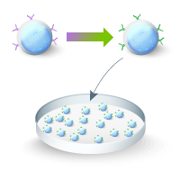
The Guide-it Long ssDNA Production System v2 employs a simple and efficient in vitro protocol for the on-demand generation of long ssDNA strands. The method involves PCR amplification of the donor template using primer pairs that include one phosphorylated oligo. Depending on whether the forward or reverse primer is phosphorylated, the resulting dsDNA PCR product is phosphorylated on either the sense or antisense strand, respectively. In the next step, the phosphorylated strand of the dsDNA is selectively digested by the nucleases Strandase A and B, generating ssDNA. This workflow is summarized by the following schematic:

Preparation of long ssDNA

Figure 1. Preparation of long ssDNA donors using the Guide-it Long ssDNA Production System v2. First, a dsDNA template (insert sequence flanked by 5' and 3' homology arms) is prepared using cloning, fusion PCR, or other related methods. The template should contain arms homologous to the target gene flanking the sequence to be inserted. Next, either of two different dsDNA PCR products is generated from the template using appropriate phosphorylated (P) primers. These PCR products serve as substrates for the strandase reaction: Strandase Mix A (purple enzyme) selectively digests the phosphorylated strand. Next, Strandase Mix B (blue enzyme) is added to finish the digestion and create ssDNA. Finally, the reaction is cleaned up to obtain ssDNA for use as an HDR template in gene knockin experiments.

Results  

Production of ssDNA templates

The Guide-it Long ssDNA Production System v2 features improvements that offer two advantages over the original (v1) ssDNA kits (Cat. # 632644, 632645):

  • A streamlined workflow involving the use of a common buffer for both strandase enzymes
  • Improved performance over the v1 kit, enabling more efficient processing of challenging templates (Figure 2, Panel A) and higher yields of ssDNA (especially for templates ranging from 500–1,500 nt)

Taking the CCR5 gene as a template for amplifying PCR products of varying lengths, we demonstrated that the kit enables the generation of ssDNA ranging from 500–5,000 nt (Figure 2, Panel B). Production of ssDNA can be confirmed on an agarose gel, with the ssDNA typically running at a lower molecular weight and yielding a band with lower intensity than the corresponding dsDNA PCR substrate.

Figure 2. Improved processing of challenging templates and production of ssDNA up to 5,000 nt with the Guide-it Long ssDNA Production System v2. Panel A. Gel image showing the dsDNA starting material (Lane 1) and sense (SS) or antisense (AS) ssDNA products (Lanes 2 and 3, respectively) for several HDR templates generated using two respective versions of the Guide-it Long ssDNA Production System (v1 or v2). The templates consisted of different inserts flanked by 5′ and 3′ homology arms targeting various genomic loci of interest and had been identified as challenging substrates for ssDNA production using the v1 kit. The dsDNA and ssDNA were analyzed via agarose gel electrophoresis using ethidium bromide as a staining agent. ssDNA products run at a smaller molecular weight than corresponding dsDNA substrates. In all cases, the v2 kit generated a cleaner band of ssDNA than the previous version of the kit, suggesting more complete and uniform digestion. Panel B. The Guide-it Long ssDNA Production System v2 enables successful production of ssDNA ranging from 500 nt to 5,000 nt in length. The gel images show dsDNA substrates of varying length (Lane 1) and corresponding sense (SS) and antisense (AS) ssDNA products (Lane 2) following enzymatic digestion and cleanup with the kit. Each ssDNA HDR template was designed to target the CCR5 gene.

Advantages of ssDNA vs. dsDNA

Reduced random integration

To compare rates of random integration for various types of donor DNA, three different types of templates (plasmid, dsDNA, and ssDNA) designed to fuse AcGFP1 to GADPH were transfected into HEK293 cells alone or in tandem with plasmids encoding Cas9 and an sgRNA targeting GADPH (Figure 3, Panel A). Three days posttransfection, frequencies of GFP-positive cells in the resulting cell populations were determined by flow cytometry (Figure 3, Panel B). The FACS results demonstrated that dsDNA yielded the highest frequency of GFP-positive cells when combined with Cas9 and sgRNA, but that a significant proportion of GFP-positive cells were also produced in the absence of Cas9/sgRNA complexes, suggesting that dsDNA donors can integrate with high efficiency at sites other than a given target site. In contrast, such random, Cas9-independent integration was not observed for ssDNA templates, which efficiently yielded GFP-positive cells only when paired with Cas9 and sgRNA.

Figure 3. Efficient editing and with negligible off-target insertion using ssDNA donor templates. Panel A. Tagging of GAPDH with a fluorescent protein (AcGFP1). An HDR template was designed to fuse the AcGFP1 coding sequence in-frame at the C-terminus of the GAPDH gene. Panel B. HEK293 cells were transfected with plasmid, dsDNA, or ssDNA forms of the AcGFP1 HDR template, with or without cotransfection of plasmids encoding for Cas9 and an sgRNA targeting the GAPDH locus. Cells were grown for three days and then analyzed by flow cytometry. While the plasmid template resulted in very little integration as demonstrated by the low percentages of fluorescent cells, the dsDNA yielded significant proportions of AcGFP1-postive cells, even in the absence of Cas9-sgRNA expression, suggesting a significant level of nonspecific integration. Both sense (S) and antisense (A) ssDNA repair templates were associated with efficient insertion of the AcGFP1 sequence in the presence but not in the absence of Cas9 and sgRNA, suggesting that insertion of the AcGFP1 sequence was highly specific.

Lower cytotoxicity

The effects of dsDNA and ssDNA donor templates on cell viability were also compared following coelectroporation with RNPs in hiPSCs and T cells, respectively (Figure 4, Panels A and B). Visualization of resulting hiPSC populations by microscopy five days postelectroporation revealed that cell counts were dramatically lower following electroporation with dsDNA, as compared to cell populations electroporated with ssDNA (Figure 5, Panel A). Similar results were obtained following electroporation of CD3+ T cells (Figure 5, Panel B). In this case, cell viability was analyzed by flow cytometry 24 hours postelectroporation.

Figure 4. Comparison of cellular toxicity induced by dsDNA and ssDNA upon coelectroporation with Cas9-sgRNA RNPs in different cell types. Panel A. Toxicity of dsDNA in hiPSCs. ChIPSC18 cells were coelectroporated with RNPs and sense or antisense ssDNA or dsDNA donor templates designed to insert AcGFP1 at the locus for the tubulin gene. The donor templates were created with 350-bp homology arms to the insertion site and were 1.5 kb in length. As seen in the images, the dsDNA donor template caused significant toxicity as compared to the ssDNA templates, resulting in lower cell counts three days after electroporation. Panel B. Toxicity of dsDNA in CD3+ T cells. Cells were electroporated with Cas9/sgRNA RNPs with or without HDR templates (dsDNA or ssDNA) designed to insert AcGFP1 at the SEC61B locus. Twenty-four hours after electroporation, cell viability was measured by flow cytometry. Electroporation of dsDNA resulted in higher levels of cell toxicity as compared to ssDNA.

Efficient insertion of an expression cassette into a safe harbor locus in hiPS cells

In a separate experiment, an HDR template was designed for the insertion of an expression cassette encoding AcGFP1 under the constitutive EF1α promoter at the AAVS1 safe harbor site (Figure 5, Panel A) in hiPSCs. The ssDNA produced from the template included 600-nt homology arms with a total length of 3,400 nt, and it was coelectroporated into hiPSCs along with RNPs targeting the AAVS1 locus. Following gene editing, FACS analysis of the resulting cell population showed that 3.64% of the cells were positive for AcGFP1 expression (Figure 6, Panel B), demonstrating efficient knockin of the sizable EF1α-AcGFP1 expression cassette.

Figure 5. Insertion of a fluorescent protein expression cassette into the AAVS1 site in hiPSCs using long ssDNA oligo as donor template. Panel A. Schematic of donor template and Cas9-sgRNA target site for insertion of AcGFP1 expression cassette at the AAVS1 locus. Panel B. FACS analysis of AcGFP1 expression in control and edited populations following electroporation.

Knockin of murine T-cell receptor α- and β-chains at the TRAC locus in primary CD3+ T cells

CAR‐T cells (chimeric antigen receptor-engineered T cells) and TCR‐T cells (T cell receptor-engineered T cells) have emerged as promising therapeutics for hematological and solid malignancies. The application of CRISPR/Cas9 in this field holds great potential, as it enables the insertion of CAR and TCR constructs into specific genomic target sites, and knockout or replacement of endogenous factors (Eyquem et al. 2017).

To demonstrate the applicability of ssDNA for immune cell therapy research, a 2.8-kb HDR template was designed for knockin of murine T-cell receptor α- and β-chains at the TRAC locus in primary human CD3+ T cells using ssDNA as a donor (Schober et al. 2019). Specifically, RNPs with sgRNAs targeting TRAC and TRBC loci, respectively, were electroporated together with the ssDNA HDR template encoding the murine T-cell receptor α- and β-chains. Knockout of both endogenous TCR genes (TRAC and TRBC) was required in order to facilitate sufficient surface expression and ensure correct pairing of the transgenic mouse receptor chains. Ten days after electroporation, flow cytometry was used to detect the successful HDR and expression of the murine TCRs (Figure 6, Panel B).

Figure 6. Knockin of murine T-cell receptor α- and β-chains at the TRAC locus in human CD3+ T cells. Panel A. CRISPR/Cas9 RNPs with sgRNAs targeting both TRAC and TRBC loci were coelectroporated with an ssDNA HDR template encoding murine T-cell receptor α- and β-chains. The HDR template was designed to include 350-nt homology arms targeting exon 1 of the TRAC locus in addition to the following elements: P2A and T2A, self-cleaving peptide inserts; TRBC mouse, mouse TCR-β constant region; TRAC mouse, mouse TCR-α constant region; pGHpA, poly-A tail. The total length of the ssDNA was 2,800 nt. Panel B. Flow cytometric analysis of resulting T-cell populations 10 days after editing using antibodies against mouse TCR-β and human TCR-α/β. Cells in the negative control population (nonedited cells) were positive for the expression of human TCRs. For cells electroporated with RNPs targeting both TRAC and TRBC loci, knockout of the endogenous TCR could be detected. In cells coelectroporated with RNPs combined with the ssDNA HDR template, expression of the murine T-cell receptor α- and β-chains could be detected.

Conclusions  

As demonstrated above, the use of ssDNA donor templates for CRISPR-based knockin applications enables more reliable editing with less nonspecific integration and lower cytotoxicity compared to dsDNA templates. However, widespread use of ssDNA has been limited for applications requiring donor templates >200 nt due to the cost and difficulty associated with producing longer ssDNA molecules. The Guide-it Long ssDNA Production System v2 addresses this limitation by enabling the on-demand production of ssDNA ranging from 500–5,000 nt in an error-free and cost-effective manner.

As shown, ssDNA produced with the Guide-it kit was associated with significantly less nonspecific integration and toxicity in comparison to dsDNA when introduced along with Cas9 RNPs into target cells and enabled efficient knockin of multi-kb constructs.

Methods  

hiPSCs used in these studies were from Cellartis Human iPS Cell Line 18 (ChiPSC18), and were cultured using Cellartis DEF-CS 500 Basal Medium with Additives.

CD3+ T cells were obtained from StemCell Technologies (Cat. # 70024) and cultured in LymphoONE T-Cell Expansion Xeno-Free Medium. Cells were activated before the gene editing experiment as described.

For electroporation experiments, sgRNA was produced using the Guide-it sgRNA In Vitro Transcription Kit and combined with Guide-it Recombinant Cas9 (Electroporation-ready) to produce Cas9 RNPs. ssDNA templates were produced using the Guide-it Long ssDNA Production System v2, according to the kit Protocol-At-A-Glance.

The different cell types were electroporated using a Neon electroporation system according to the following guidelines:

hiPSCs: 1.5 x 10⁵ cells were electroporated in Buffer R with 1 µg of ssDNA or dsDNA in either the presence or absence of 2.25 µg of Cas9 nuclease combined with 0.45 µg of sgRNA. The electroporation conditions were as follows: pulse voltage / pulse width / pulse number = 1100 V / 20 ms / 2 pulses

CD3+ T cells: 3 x 10⁵ cells were electroporated in Buffer T with 0.75 µg of ssDNA in the presence of 1.68 µg of Cas9 nuclease combined with 0.343 µg of sgRNA. The electroporation conditions were as follows: pulse voltage / pulse width / pulse number = 1600 V / 10 ms / 3 pulses

Cytotoxicity of hiPSCs was assessed 48 hr after electroporation by microscopy.

Cytotoxicity of T cells was assessed 24 hr after electroporation by flow cytometry using eBioscience Fixable Viability Dye eFluor 660 (Thermo Scientific Cat# 65-0864-14). Successful KI was detected using the following antibodies: PE anti-human TCR-α/β (Biolegend Cat# 306707) and FITC anti-murine TCR β-chain–APC (Biolegend Cat# 109205).

For the experiments involving HEK293 cells, we used the Guide-it CRISPR/Cas9 System, consisting of a plasmid expressing Cas9 and the sgRNA. The Cas9-sgRNA plasmid was transfected along with the donor templates using Xfect Transfection Reagent. Again, long ssDNA was produced according to the user manual for the Guide-it Long ssDNA Production System v2, and 1 µg of ssDNA or dsDNA was cotransfected along with the Cas9-sgRNA expression plasmid. The transfected cells were subjected to flow cytometry analysis three days posttransfection.

References  

Chen et al., High-frequency genome editing using ssDNA oligonucleotides with zinc-finger nucleases. Nat. Meth. 8, 753–755 (2011).

Eyquem et al., Targeting a CAR to the TRAC locus with CRISPR/Cas9 enhances tumour rejection. Nature 543(7643), 113–117 (2017).

Roth et al., Reprogramming human T cell function and specificity with nonviral genome targeting. Nature 559(7714), 405–409 (2018).

Schober et al., Orthotopic replacement of T-cell receptor a- and b- chains with preservation of near-physiological T-cell function. Nature Biomedical Engineering 543(12), 974–984 (2019).

Related Products

Cat. # Product Size Price License Quantity Details
632666 Guide-it™ Long ssDNA Production System v2 50 Rxns USD $605.00

License Statement

ID Number  
M54 This product is covered by the claims of U.S. Patent Nos. 7,704,713 and its foreign counterparts. 

The Guide-it Long ssDNA Production System v2 includes all the reagents and materials necessary for on-demand preparation of single-stranded DNA (ssDNA) repair templates of any sequence up to 5,000 nt in length for engineering gene knockins using CRISPR/Cas9 technology or other genome editing tools. It has been demonstrated that ssDNA offers two key advantages over dsDNA templates for precise genome editing applications: greatly reduced toxicity and a much lower likelihood of random or off-target integration. The kit employs a simple and fast method that involves conversion of a dsDNA PCR product into ssDNA via selective digestion of either the sense or the antisense strand. Following digestion, ssDNA products are purified using silica membrane spin columns included with the kit.

Cat. # 632666 includes sufficient quantities of reagents and materials for preparation of 50 ssDNA templates.

Notice to purchaser

Our products are to be used for Research Use Only. They may not be used for any other purpose, including, but not limited to, use in humans, therapeutic or diagnostic use, or commercial use of any kind. Our products may not be transferred to third parties, resold, modified for resale, or used to manufacture commercial products or to provide a service to third parties without our prior written approval.

Documents Components Image Data

Back

Improved processing of challenging templates and production of ssDNA up to 5,000 nt with the Guide-it Long ssDNA Production System v2.

Improved processing of challenging templates and production of ssDNA up to 5,000 nt with the Guide-it Long ssDNA Production System v2.

Improved processing of challenging templates and production of ssDNA up to 5,000 nt with the Guide-it Long ssDNA Production System v2. The Guide-it Long ssDNA Production System v2 enables successful production of ssDNA ranging from 500 nt to 5,000 nt in length. The gel images show dsDNA substrates of varying length (Lane 1) and corresponding sense (SS) and antisense (AS) ssDNA products (Lane 2) following enzymatic digestion and cleanup with the kit. Each ssDNA HDR template was designed to target the CCR5 gene.

Back

Efficient editing and with negligible off-target insertion using ssDNA donor templates.

Efficient editing and with negligible off-target insertion using ssDNA donor templates.

Efficient editing and with negligible off-target insertion using ssDNA donor templates. Panel A. Tagging of GAPDH with a fluorescent protein (AcGFP1). An HDR template was designed to fuse the AcGFP1 coding sequence in-frame at the C-terminus of the GAPDH gene. Panel B. HEK293 cells were transfected with plasmid, dsDNA, or ssDNA forms of the AcGFP1 HDR template, with or without co-transfection of plasmids encoding for Cas9 and an sgRNA targeting the GAPDH locus. Cells were grown for three days and then analyzed by flow cytometry. While the plasmid template resulted in very little integration as demonstrated by the low percentages of fluorescent cells, the dsDNA yielded significant proportions of AcGFP1-postive cells, even in the absence of Cas9-sgRNA expression, suggesting a significant level of non-specific integration. Both sense (S) and antisense (A) ssDNA repair templates were associated with efficient insertion of the AcGFP1 sequence in the presence but not in the absence of Cas9 and sgRNA, suggesting that insertion of the AcGFP1 sequence was highly specific.

Back

Knockin of murine T-cell receptor α- and β-chains at the TRAC locus in human CD3+ T cells.

Knockin of murine T-cell receptor α- and β-chains at the TRAC locus in human CD3+ T cells.

Knockin of murine T-cell receptor α- and β-chains at the TRAC locus in human CD3+ T cells. Panel A. CRISPR/Cas9 RNPs with sgRNAs targeting both TRAC and TRBC loci were coelectroporated with an ssDNA HDR template encoding murine T-cell receptor α- and β-chains. The HDR template was designed to include 350-nt homology arms targeting exon 1 of the TRAC locus in addition to the following elements: P2A and T2A, self-cleaving peptide inserts; TRBC mouse, mouse TCR-β constant region; TRAC mouse, mouse TCR-α constant region; pGHpA, poly-A tail. The total length of the ssDNA was 2,800 nt. Panel B. Flow cytometric analysis of resulting T-cell populations 10 days after editing using antibodies against mouse TCR-β and human TCR-α/β. Cells in the negative control population (nonedited cells) were positive for the expression of human TCRs. For cells electroporated with RNPs targeting both TRAC and TRBC loci, knockout of the endogenous TCR could be detected. In cells coelectroporated with RNPs combined with the ssDNA HDR template, expression of the murine T-cell receptor α- and β-chains could be detected.

Back

632666: Guide-it Long ssDNA Production System v2

632666: Guide-it Long ssDNA Production System v2


Choose an HDR template format

Watch a webinar on how to choose the right HDR template for knockin experiments.

Homology-directed repair FAQs

Maximize your likelihood for success by reading these FAQs compiled by our R&D team.

Gene editing of T cells and HSCs

Follow these protocols developed by our R&D team for easy CRISPR/Cas-based gene editing of immune cells.

Site-specific gene knockins using long ssDNA

Gene knockins with long ssDNA sequences produced using Guide-it Long ssDNA Production System v2.

CRISPR/Cas9-mediated knockins in iPS cells

Data demonstrating efficient genome editing of iPS cells using HDR templates generated with the Guide-it Long ssDNA Production System.

CRISPR takes a giant step towards the clinic

Learn about the development of a powerful new method for reprogramming T cells.

Mouse CRISPR knockin protocol

Access a customer-developed protocol for precise genome editing in mouse embryos.

Electroporation-grade Cas9 for editing in diverse cell types

Our Cas9 performs highly efficient gene editing, including in iPS and hematopoietic stem cells.

Screening for effective guide RNAs

Before delivering sgRNA to your cells, use a novel in vitro assay to get accurate predictions of sgRNA cleavage efficiency.

Tag an endogenous gene with AcGFP1 in iPS cells

Workflow for tagging endogenous genes to generate reporter lines.

Tag an endogenous gene with a myc tag in iPS cells

Workflow for tagging endogenous genes with an epitope tag.

Efficient SNP engineering

Learn about a simple assay for sensitive detection of single-nucleotide substitutions in bulk-edited or clonal cell populations.

Takara Bio USA, Inc.
United States/Canada: +1.800.662.2566 • Asia Pacific: +1.650.919.7300 • Europe: +33.(0)1.3904.6880 • Japan: +81.(0)77.565.6999
FOR RESEARCH USE ONLY. NOT FOR USE IN DIAGNOSTIC PROCEDURES. © 2025 Takara Bio Inc. All Rights Reserved. All trademarks are the property of Takara Bio Inc. or its affiliate(s) in the U.S. and/or other countries or their respective owners. Certain trademarks may not be registered in all jurisdictions. Additional product, intellectual property, and restricted use information is available at takarabio.com.

Takara Bio

Takara Bio USA, Inc. provides kits, reagents, instruments, and services that help researchers explore questions about gene discovery, regulation, and function. As a member of the Takara Bio Group, Takara Bio USA is part of a company that holds a leadership position in the global market and is committed to improving the human condition through biotechnology. Our mission is to develop high-quality innovative tools and services to accelerate discovery.

FOR RESEARCH USE ONLY. NOT FOR USE IN DIAGNOSTIC PROCEDURES (EXCEPT AS SPECIFICALLY NOTED).

Support
  • Contact us
  • Technical support
  • Customer service
  • Shipping & delivery
  • Sales
  • Feedback
Products
  • New products
  • Special offers
  • Instrument & reagent services
Learning centers
  • NGS
  • Gene function
  • Stem cell research
  • Protein research
  • PCR
  • Cloning
  • Nucleic acid purification
About
  • Our brands
  • Careers
  • Events
  • Blog
  • Need help?
  • Announcements
  • Quality and compliance
  • That's Good Science!
Facebook Twitter  LinkedIn

logo strip white

©2025 Takara Bio Inc. All Rights Reserved.

Region - North America Privacy Policy Terms and Conditions Terms of Use

Top


  • COVID-19 research
  • Viral detection with qPCR
  • SARS-CoV-2 pseudovirus
  • Human ACE2 stable cell line
  • Viral RNA isolation
  • Viral and host sequencing
  • Vaccine development
  • CRISPR screening
  • Drug discovery
  • Immune profiling
  • Publications
  • Next-generation sequencing
  • Spatial omics
  • RNA-seq
  • DNA-seq
  • Single-cell NGS automation
  • Reproductive health
  • Bioinformatics tools
  • Immune profiling
  • Real-time PCR
  • Great value master mixes
  • Signature enzymes
  • High-throughput real-time PCR solutions
  • Detection assays
  • References, standards, and buffers
  • Stem cell research
  • Media, differentiation kits, and matrices
  • Stem cells and stem cell-derived cells
  • mRNA and cDNA synthesis
  • In vitro transcription
  • cDNA synthesis kits
  • Reverse transcriptases
  • RACE kits
  • Purified cDNA & genomic DNA
  • Purified total RNA and mRNA
  • PCR
  • Most popular polymerases
  • High-yield PCR
  • High-fidelity PCR
  • GC rich PCR
  • PCR master mixes
  • Cloning
  • In-Fusion seamless cloning
  • Competent cells
  • Ligation kits
  • Restriction enzymes
  • Nucleic acid purification
  • Automated platforms
  • Plasmid purification kits
  • Genomic DNA purification kits
  • DNA cleanup kits
  • RNA purification kits
  • Gene function
  • Gene editing
  • Viral transduction
  • Fluorescent proteins
  • T-cell transduction and culture
  • Tet-inducible expression systems
  • Transfection reagents
  • Cell biology assays
  • Protein research
  • Purification products
  • Two-hybrid and one-hybrid systems
  • Mass spectrometry reagents
  • Antibodies and ELISAs
  • Primary antibodies and ELISAs by research area
  • Fluorescent protein antibodies
  • New products
  • Special offers
  • OEM
  • Portfolio
  • Process
  • Facilities
  • Request samples
  • FAQs
  • Instrument services
  • Apollo services
  • ICELL8 services
  • SmartChip ND system services
  • Gene and cell therapy manufacturing services
  • Services
  • Facilities
  • Our process
  • Resources
  • Customer service
  • Sales
  • Make an appointment with your sales rep
  • Shipping & delivery
  • Technical support
  • Feedback
  • Online tools
  • GoStix Plus FAQs
  • Partnering & Licensing
  • Vector information
  • Vector document overview
  • Vector document finder
Takara Bio's award-winning GMP-compliant manufacturing facility in Kusatsu, Shiga, Japan.

Partner with Takara Bio!

Takara Bio is proud to offer GMP-grade manufacturing capabilities at our award-winning facility in Kusatsu, Shiga, Japan.

  • Automation systems
  • Shasta Single Cell System introduction
  • SmartChip Real-Time PCR System introduction
  • ICELL8 introduction
  • Next-generation sequencing
  • RNA-seq
  • Technical notes
  • Technology and application overviews
  • FAQs and tips
  • DNA-seq protocols
  • Bioinformatics resources
  • Webinars
  • Spatial biology
  • Real-time PCR
  • Download qPCR resources
  • Overview
  • Reaction size guidelines
  • Guest webinar: extraction-free SARS-CoV-2 detection
  • Technical notes
  • Nucleic acid purification
  • Nucleic acid extraction webinars
  • Product demonstration videos
  • Product finder
  • Plasmid kit selection guide
  • RNA purification kit finder
  • mRNA and cDNA synthesis
  • mRNA synthesis
  • cDNA synthesis
  • PCR
  • Citations
  • PCR selection guide
  • Technical notes
  • FAQ
  • Cloning
  • Automated In-Fusion Cloning
  • In-Fusion Cloning general information
  • Primer design and other tools
  • In‑Fusion Cloning tips and FAQs
  • Applications and technical notes
  • Stem cell research
  • Overview
  • Protocols
  • Technical notes
  • Gene function
  • Gene editing
  • Viral transduction
  • T-cell transduction and culture
  • Inducible systems
  • Cell biology assays
  • Protein research
  • Capturem technology
  • Antibody immunoprecipitation
  • His-tag purification
  • Other tag purification
  • Expression systems
  • Antibodies and ELISA
  • Molecular diagnostics
  • Interview: adapting to change with Takara Bio
  • Applications
  • Solutions
  • Partnering
  • Contact us
  • mRNA and protein therapeutics
  • Characterizing the viral genome and host response
  • Identifying and cloning protein targets
  • Expressing and purifying protein targets
  • Immunizing mice and optimizing vaccines
  • Pathogen detection
  • Sample prep
  • Detection methods
  • Identification and characterization
  • SARS-CoV-2
  • Antibiotic-resistant bacteria
  • Food crop pathogens
  • Waterborne disease outbreaks
  • Viral-induced cancer
  • Immunotherapy research
  • T-cell therapy
  • Antibody therapeutics
  • T-cell receptor profiling
  • TBI initiatives in cancer therapy
  • Cancer research
  • Kickstart your cancer research with long-read sequencing
  • Sample prep from FFPE tissue
  • Sample prep from plasma
  • Cancer biomarker quantification
  • Single cancer cell analysis
  • Cancer transcriptome analysis
  • Cancer genomics and epigenomics
  • HLA typing in cancer
  • Gene editing for cancer therapy/drug discovery
  • Alzheimer's disease research
  • Antibody engineering
  • Sample prep from FFPE tissue
  • Single-cell sequencing
  • Reproductive health technologies
  • Embgenix FAQs
  • Preimplantation genetic testing
  • ESM partnership program
  • ESM Collection Kit forms
  • Infectious diseases
  • Develop vaccines for HIV
Create a web account with us

Log in to enjoy additional benefits

Want to save this information?

An account with takarabio.com entitles you to extra features such as:

•  Creating and saving shopping carts
•  Keeping a list of your products of interest
•  Saving all of your favorite pages on the site*
•  Accessing restricted content

*Save favorites by clicking the star () in the top right corner of each page while you're logged in.

Create an account to get started

  • BioView blog
  • Automation
  • Cancer research
  • Career spotlights
  • Current events
  • Customer stories
  • Gene editing
  • Research news
  • Single-cell analysis
  • Stem cell research
  • Tips and troubleshooting
  • Women in STEM
  • That's Good Support!
  • About our blog
  • That's Good Science!
  • SMART-Seq Pro Biomarker Discovery Contest
  • DNA extraction educational activity
  • That's Good Science Podcast
  • Season one
  • Season two
  • Season three
  • Our brands
  • Our history
  • In the news
  • Events
  • Biomarker discovery events
  • Calendar
  • Conferences
  • Speak with us
  • Careers
  • Company benefits
  • Trademarks
  • License statements
  • Quality statement
  • HQ-grade reagents
  • International Contacts by Region
  • United States and Canada
  • China
  • Japan
  • Korea
  • Europe
  • India
  • Affiliates & distributors
  • Need help?
  • Privacy request
  • Website FAQs

That's GOOD Science!

What does it take to generate good science? Careful planning, dedicated researchers, and the right tools. At Takara Bio, we thoughtfully develop exceptional products to tackle your most challenging research problems, and have an expert team of technical support professionals to help you along the way, all at superior value.

Explore what makes good science possible

 Customer Login
 View Cart (0)
Takara Bio
  • Home
  • Products
  • Services & Support
  • Learning centers
  • APPLICATIONS
  • About
  • Contact Us
  •  Customer Login
  • Register
  •  View Cart (0)

Takara Bio USA, Inc. provides kits, reagents, instruments, and services that help researchers explore questions about gene discovery, regulation, and function. As a member of the Takara Bio Group, Takara Bio USA is part of a company that holds a leadership position in the global market and is committed to improving the human condition through biotechnology. Our mission is to develop high-quality innovative tools and services to accelerate discovery.

FOR RESEARCH USE ONLY. NOT FOR USE IN DIAGNOSTIC PROCEDURES (EXCEPT AS SPECIFICALLY NOTED).

Clontech, TaKaRa, cellartis

  • Products
  • COVID-19 research
  • Next-generation sequencing
  • Real-time PCR
  • Stem cell research
  • mRNA and cDNA synthesis
  • PCR
  • Cloning
  • Nucleic acid purification
  • Gene function
  • Protein research
  • Antibodies and ELISA
  • New products
  • Special offers
  • COVID-19 research
  • Viral detection with qPCR
  • SARS-CoV-2 pseudovirus
  • Human ACE2 stable cell line
  • Viral RNA isolation
  • Viral and host sequencing
  • Vaccine development
  • CRISPR screening
  • Drug discovery
  • Immune profiling
  • Publications
  • Next-generation sequencing
  • Spatial omics
  • RNA-seq
  • DNA-seq
  • Single-cell NGS automation
  • Reproductive health
  • Bioinformatics tools
  • Immune profiling
  • Real-time PCR
  • Great value master mixes
  • Signature enzymes
  • High-throughput real-time PCR solutions
  • Detection assays
  • References, standards, and buffers
  • Stem cell research
  • Media, differentiation kits, and matrices
  • Stem cells and stem cell-derived cells
  • mRNA and cDNA synthesis
  • In vitro transcription
  • cDNA synthesis kits
  • Reverse transcriptases
  • RACE kits
  • Purified cDNA & genomic DNA
  • Purified total RNA and mRNA
  • PCR
  • Most popular polymerases
  • High-yield PCR
  • High-fidelity PCR
  • GC rich PCR
  • PCR master mixes
  • Cloning
  • In-Fusion seamless cloning
  • Competent cells
  • Ligation kits
  • Restriction enzymes
  • Nucleic acid purification
  • Automated platforms
  • Plasmid purification kits
  • Genomic DNA purification kits
  • DNA cleanup kits
  • RNA purification kits
  • Gene function
  • Gene editing
  • Viral transduction
  • Fluorescent proteins
  • T-cell transduction and culture
  • Tet-inducible expression systems
  • Transfection reagents
  • Cell biology assays
  • Protein research
  • Purification products
  • Two-hybrid and one-hybrid systems
  • Mass spectrometry reagents
  • Antibodies and ELISA
  • Primary antibodies and ELISAs by research area
  • Fluorescent protein antibodies
  • Services & Support
  • OEM
  • Instrument services
  • Gene and cell therapy manufacturing
  • Customer service
  • Sales
  • Shipping & delivery
  • Technical support
  • Feedback
  • Online tools
  • Partnering & Licensing
  • Vector information
  • OEM
  • Portfolio
  • Process
  • Facilities
  • Request samples
  • FAQs
  • Instrument services
  • Apollo services
  • ICELL8 services
  • SmartChip ND system services
  • Gene and cell therapy manufacturing
  • Services
  • Facilities
  • Our process
  • Resources
  • Sales
  • Make an appointment with your sales rep
  • Online tools
  • GoStix Plus FAQs
  • Vector information
  • Vector document overview
  • Vector document finder
  • Learning centers
  • Automation systems
  • Next-generation sequencing
  • Spatial biology
  • Real-time PCR
  • Nucleic acid purification
  • mRNA and cDNA synthesis
  • PCR
  • Cloning
  • Stem cell research
  • Gene function
  • Protein research
  • Antibodies and ELISA
  • Automation systems
  • Shasta Single Cell System introduction
  • SmartChip Real-Time PCR System introduction
  • ICELL8 introduction
  • Next-generation sequencing
  • RNA-seq
  • Technical notes
  • Technology and application overviews
  • FAQs and tips
  • DNA-seq protocols
  • Bioinformatics resources
  • Webinars
  • Real-time PCR
  • Download qPCR resources
  • Overview
  • Reaction size guidelines
  • Guest webinar: extraction-free SARS-CoV-2 detection
  • Technical notes
  • Nucleic acid purification
  • Nucleic acid extraction webinars
  • Product demonstration videos
  • Product finder
  • Plasmid kit selection guide
  • RNA purification kit finder
  • mRNA and cDNA synthesis
  • mRNA synthesis
  • cDNA synthesis
  • PCR
  • Citations
  • PCR selection guide
  • Technical notes
  • FAQ
  • Cloning
  • Automated In-Fusion Cloning
  • In-Fusion Cloning general information
  • Primer design and other tools
  • In‑Fusion Cloning tips and FAQs
  • Applications and technical notes
  • Stem cell research
  • Overview
  • Protocols
  • Technical notes
  • Gene function
  • Gene editing
  • Viral transduction
  • T-cell transduction and culture
  • Inducible systems
  • Cell biology assays
  • Protein research
  • Capturem technology
  • Antibody immunoprecipitation
  • His-tag purification
  • Other tag purification
  • Expression systems
  • APPLICATIONS
  • Molecular diagnostics
  • mRNA and protein therapeutics
  • Pathogen detection
  • Immunotherapy research
  • Cancer research
  • Alzheimer's disease research
  • Reproductive health technologies
  • Infectious diseases
  • Molecular diagnostics
  • Interview: adapting to change with Takara Bio
  • Applications
  • Solutions
  • Partnering
  • Contact us
  • mRNA and protein therapeutics
  • Characterizing the viral genome and host response
  • Identifying and cloning protein targets
  • Expressing and purifying protein targets
  • Immunizing mice and optimizing vaccines
  • Pathogen detection
  • Sample prep
  • Detection methods
  • Identification and characterization
  • SARS-CoV-2
  • Antibiotic-resistant bacteria
  • Food crop pathogens
  • Waterborne disease outbreaks
  • Viral-induced cancer
  • Immunotherapy research
  • T-cell therapy
  • Antibody therapeutics
  • T-cell receptor profiling
  • TBI initiatives in cancer therapy
  • Cancer research
  • Kickstart your cancer research with long-read sequencing
  • Sample prep from FFPE tissue
  • Sample prep from plasma
  • Cancer biomarker quantification
  • Single cancer cell analysis
  • Cancer transcriptome analysis
  • Cancer genomics and epigenomics
  • HLA typing in cancer
  • Gene editing for cancer therapy/drug discovery
  • Alzheimer's disease research
  • Antibody engineering
  • Sample prep from FFPE tissue
  • Single-cell sequencing
  • Reproductive health technologies
  • Embgenix FAQs
  • Preimplantation genetic testing
  • ESM partnership program
  • ESM Collection Kit forms
  • Infectious diseases
  • Develop vaccines for HIV
  • About
  • BioView blog
  • That's Good Science!
  • Our brands
  • Our history
  • In the news
  • Events
  • Careers
  • Trademarks
  • License statements
  • Quality and compliance
  • HQ-grade reagents
  • International Contacts by Region
  • Need help?
  • Website FAQs
  • BioView blog
  • Automation
  • Cancer research
  • Career spotlights
  • Current events
  • Customer stories
  • Gene editing
  • Research news
  • Single-cell analysis
  • Stem cell research
  • Tips and troubleshooting
  • Women in STEM
  • That's Good Support!
  • About our blog
  • That's Good Science!
  • SMART-Seq Pro Biomarker Discovery Contest
  • DNA extraction educational activity
  • That's Good Science Podcast
  • Season one
  • Season two
  • Season three
  • Events
  • Biomarker discovery events
  • Calendar
  • Conferences
  • Speak with us
  • Careers
  • Company benefits
  • International Contacts by Region
  • United States and Canada
  • China
  • Japan
  • Korea
  • Europe
  • India
  • Affiliates & distributors
  • Need help?
  • Privacy request
Takara Bio
  • Products
  • Services & Support
  • Learning centers
  • APPLICATIONS
  • About
  • Contact Us