We use cookies to improve your browsing experience and provide meaningful content. Read our cookie policy. Accept
  •  Customer Login
  • Register
  •  View Cart (0)
  •  Customer Login
  • Register
  •  View Cart (0)

Takara Bio
  • Products
  • Services & Support
  • Learning centers
  • APPLICATIONS
  • About
  • Contact Us

Clontech Takara Cellartis

Close

  • ‹ Back to Far-red fluorescent proteins
  • E2-Crimson fluorescent protein
  • HcRed1 fluorescent protein
  • mRaspberry fluorescent protein
  • mPlum fluorescent protein
Selection guides Fluorescent protein quick guide
Tips and FAQs
In‑Fusion Cloning tips and FAQs
Home › Products › Gene function › Fluorescent proteins › Fluorescent protein plasmids › Far-red fluorescent proteins › E2-Crimson fluorescent protein

Fluorescent protein plasmids

  • Cyan and green fluorescent proteins
    • AcGFP1 fluorescent protein
    • ZsGreen1 fluorescent protein
    • GFP & GFPuv fluorescent proteins
    • AmCyan1 fluorescent protein
  • Red fluorescent proteins
    • mCherry fluorescent protein
    • DsRed-Monomer fluorescent protein
    • DsRed2 fluorescent protein
    • DsRed-Express and DsRed-Express2 fluorescent proteins
    • tdTomato fluorescent protein
    • AsRed2 fluorescent protein
    • mStrawberry fluorescent protein
  • Far-red fluorescent proteins
    • E2-Crimson fluorescent protein
    • HcRed1 fluorescent protein
    • mRaspberry fluorescent protein
    • mPlum fluorescent protein
  • Orange and yellow fluorescent proteins
    • mOrange2 fluorescent protein
    • mBanana fluorescent protein
    • ZsYellow1 fluorescent protein
  • Photoactivatable and photoswitchable proteins
    • Dendra2 fluorescent protein
    • Timer fluorescent protein
    • PAmCherry fluorescent protein
Need help?
Contact Sales
Selection guides Fluorescent protein quick guide
Tips and FAQs
In‑Fusion Cloning tips and FAQs

E2-Crimson fluorescent protein

E2-Crimson fluorescent protein vector

Living Colors E2-Crimson is a bright far-red fluorescent protein that was designed for in vivo applications involving sensitive cells, such as primary cells and stem cells. E2-Crimson was derived from DsRed-Express2 and retains its rapid maturation (half-time of 26 minutes at 37°C), high photostability, high solubility, and low cytotoxicity (Strack et al. 2009).

Cat. # Product Size Price License Quantity Details
632556 pCMV-E2-Crimson Vector 20 ug Inquire for Quotation *

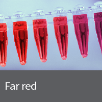
The mammalian CMV promoter in pCMV-E2-Crimson allows the constitutive expression of the far-red Living Colors fluorescent protein E2-Crimson, driven by the constitutive immediate early promoter of cytomegalovirus (CMV). pCMV-E2-Crimson does not contain an MCS. It can be used for labeling purposes or as a cotransfection marker to determine transfection efficiency. If required, stable transfectants can be selected using G418.

Notice to purchaser

Our products are to be used for Research Use Only. They may not be used for any other purpose, including, but not limited to, use in humans, therapeutic or diagnostic use, or commercial use of any kind. Our products may not be transferred to third parties, resold, modified for resale, or used to manufacture commercial products or to provide a service to third parties without our prior written approval.

Documents Components You May Also Like
632555 pE2-Crimson-C1 Vector 20 ug Inquire for Quotation *

pE2-Crimson-C1 is a mammalian expression vector designed to express a protein of interest fused to the C-terminus of E2-Crimson, our brightest and fastest-maturing far-red Living Colors fluorescent protein. E2-Crimson was derived from DsRed-Express2, and retains its fast maturation, high photostability, increased solubility, and low cytotoxicity. The unmodified vector can be used to express E2-Crimson in mammalian cells. If required, stable transfectants can be selected using G418.

Notice to purchaser

Our products are to be used for Research Use Only. They may not be used for any other purpose, including, but not limited to, use in humans, therapeutic or diagnostic use, or commercial use of any kind. Our products may not be transferred to third parties, resold, modified for resale, or used to manufacture commercial products or to provide a service to third parties without our prior written approval.

Documents Components You May Also Like Image Data

Back

Schematic for fluorescent fusion protein vectors

Schematic for fluorescent fusion protein vectors

Schematic for fluorescent fusion protein vectors. Panel A. N-terminal fluorescent protein (FP) vector. Panel B. C-terminal fluorescent protein vector.

Back

E2-Crimson is useful for confocal and STED (stimulated emission depletion) microscopy

E2-Crimson is useful for confocal and STED (stimulated emission depletion) microscopy
E2-Crimson is useful for confocal and STED (stimulated emission depletion) microscopy. The mammalian ER was imaged by conventional confocal microscopy (left) or by STED microscopy (right) with 635 nm excitation and a STED wavelength of 760 nm. The scale bar is 1 μm.
632554 pE2-Crimson-N1 Vector 20 ug Inquire for Quotation *

pE2-Crimson-N1 is a mammalian expression vector designed to express a protein of interest fused to the N-terminus of E2-Crimson, our brightest and fastest-maturing far-red Living Colors fluorescent protein. E2-Crimson was derived from DsRed-Express2, and retains its fast maturation, high photostability, increased solubility, and low cytotoxicity. The unmodified vector can be used to express E2-Crimson in mammalian cells. If required, stable transfectants can be selected using G418.

Notice to purchaser

Our products are to be used for Research Use Only. They may not be used for any other purpose, including, but not limited to, use in humans, therapeutic or diagnostic use, or commercial use of any kind. Our products may not be transferred to third parties, resold, modified for resale, or used to manufacture commercial products or to provide a service to third parties without our prior written approval.

Documents Components You May Also Like Image Data

Back

Schematic for fluorescent fusion protein vectors

Schematic for fluorescent fusion protein vectors

Schematic for fluorescent fusion protein vectors. Panel A. N-terminal fluorescent protein (FP) vector. Panel B. C-terminal fluorescent protein vector.

Back

E2-Crimson is useful for confocal and STED (stimulated emission depletion) microscopy

E2-Crimson is useful for confocal and STED (stimulated emission depletion) microscopy
E2-Crimson is useful for confocal and STED (stimulated emission depletion) microscopy. The mammalian ER was imaged by conventional confocal microscopy (left) or by STED microscopy (right) with 635 nm excitation and a STED wavelength of 760 nm. The scale bar is 1 μm.
632553 pE2-Crimson Vector 20 ug Inquire for Quotation *

pE2-Crimson is a prokaryotic expression vector which encodes E2-Crimson, our brightest and fastest-maturing far-red Living Colors fluorescent protein. E2-Crimson was derived from DsRed-Express2, and retains its fast maturation, high photostability, increased solubility, and low cytotoxicity. pE2-Crimson is primarily intended to serve as a source of E2-Crimson cDNA. The flanking MCS regions make it possible to excise the E2-Crimson coding sequence and insert it into other expression vectors of choice. The vector can also be used in bacteria to produce E2-Crimson protein.

Notice to purchaser

Our products are to be used for Research Use Only. They may not be used for any other purpose, including, but not limited to, use in humans, therapeutic or diagnostic use, or commercial use of any kind. Our products may not be transferred to third parties, resold, modified for resale, or used to manufacture commercial products or to provide a service to third parties without our prior written approval.

Documents Components You May Also Like Image Data

Back

Basic fluorescent protein vector map

Basic fluorescent protein vector map

Basic fluorescent protein vector map. Use this bacterial expression vector as a source of the fluorescent protein gene. Note: There is a stop codon at the 5' end of the 3' MCS. The 3' MCS should not be used for cloning.

Back

E2-Crimson is useful for confocal and STED (stimulated emission depletion) microscopy

E2-Crimson is useful for confocal and STED (stimulated emission depletion) microscopy
E2-Crimson is useful for confocal and STED (stimulated emission depletion) microscopy. The mammalian ER was imaged by conventional confocal microscopy (left) or by STED microscopy (right) with 635 nm excitation and a STED wavelength of 760 nm. The scale bar is 1 μm.
631981 pEF1alpha-E2-Crimson Vector 10 ug Inquire for Quotation *

pEF1α-E2-Crimson is a mammalian expression vector that constitutively expresses the far-red fluorescent protein E2-Crimson, even after stable integration of the vector into the host cell genome. Stable, constitutive expression of E2-Crimson is driven by the human elongation factor 1 alpha (EF1α) promoter, which allows the protein to be expressed without the transgene silencing associated with CMV promoters. The vector, which lacks an MCS, is designed to be used for cell labeling or as a marker of transfection efficiency.

Notice to purchaser

Our products are to be used for Research Use Only. They may not be used for any other purpose, including, but not limited to, use in humans, therapeutic or diagnostic use, or commercial use of any kind. Our products may not be transferred to third parties, resold, modified for resale, or used to manufacture commercial products or to provide a service to third parties without our prior written approval.

Documents Components You May Also Like Image Data

Back

Map schematic of the plasmid choices that are available carrying the EF1-alpha promoter

Map schematic of the plasmid choices that are available carrying the EF1-alpha promoter

Map schematic of the plasmid choices that are available carrying the EF1-alpha promoter. IRES: internal ribosome entry sequence; FP1: fluorescent protein (AcGFP1, DsRed-Monomer, or mCherry); FP2: fluorescent protein (mCherry or ZsGreen1); MCS: multiple cloning site.

*You must be logged in to a Purchasing Account in order to purchase these products online, since the purchase of these products may be restricted depending on your account type. Researchers at not-for-profit accounts receive a limited use license with their purchase of the product. Researchers at for-profit accounts must obtain a license prior to purchase. For details please contact licensing@takarabio.com.

Antibodies to GFP variants, mCherry and other fluorescent proteins Fluorescent protein antibodies
Selection guides Fluorescent lentiviral particles

Overview

  • Far-red fluorescent protein
  • E2-Crimson fluorescent protein excitation and emission maxima: 611 and 646 nm, respectively
  • Several of our red fluorescent protein antibodies strongly detect E2-Crimson

More Information

Applications

  • Applications involving sensitive cells, such as primary cells and stem cells
  • Fusions
  • Protein localization studies
  • General reporter for mammalian cells
  • Monitoring transfection efficiencies
  • In vivo imaging
References

Strack, R. L. et al. A rapidly maturing far-red derivative of DsRed-Express2 for whole-cell labeling. Biochemistry 48, 8279–8281 (2009).

Additional product information

Please see the product's Certificate of Analysis for information about storage conditions, product components, and technical specifications. Please see the Kit Components List to determine kit components. Certificates of Analysis and Kit Components Lists are located under the Documents tab.


Fluorescent protein related links

  • Fluorescent protein quick guide
  • Fluorescent protein antibodies
  • GFP plasmids
  • mCherry plasmids
  • tdTomato plasmids
  • Orange/yellow fluorescent proteins
  • Photoactivatable/photoswitchable fluorescent proteins
  • Flow cytometer calibration beads
  • Purified recombinant fluorescent proteins
  • Reporting promoter activity
  • MicroRNA activity reporter
  • Labeling organelles and cellular compartments
  • Ready-to-use lentivirus
  • Lentivirus vectors with fluorescent proteins
  • Retrovirus vectors with fluorescent proteins
  • Adenovirus vectors with fluorescent proteins
  • Tetracycline-inducible systems with fluorescent proteins

Takara Bio USA, Inc.
United States/Canada: +1.800.662.2566 • Asia Pacific: +1.650.919.7300 • Europe: +33.(0)1.3904.6880 • Japan: +81.(0)77.565.6999
FOR RESEARCH USE ONLY. NOT FOR USE IN DIAGNOSTIC PROCEDURES. © 2025 Takara Bio Inc. All Rights Reserved. All trademarks are the property of Takara Bio Inc. or its affiliate(s) in the U.S. and/or other countries or their respective owners. Certain trademarks may not be registered in all jurisdictions. Additional product, intellectual property, and restricted use information is available at takarabio.com.

Takara Bio

Takara Bio USA, Inc. provides kits, reagents, instruments, and services that help researchers explore questions about gene discovery, regulation, and function. As a member of the Takara Bio Group, Takara Bio USA is part of a company that holds a leadership position in the global market and is committed to improving the human condition through biotechnology. Our mission is to develop high-quality innovative tools and services to accelerate discovery.

FOR RESEARCH USE ONLY. NOT FOR USE IN DIAGNOSTIC PROCEDURES (EXCEPT AS SPECIFICALLY NOTED).

Support
  • Contact us
  • Technical support
  • Customer service
  • Shipping & delivery
  • Sales
  • Feedback
Products
  • New products
  • Special offers
  • Instrument & reagent services
Learning centers
  • NGS
  • Gene function
  • Stem cell research
  • Protein research
  • PCR
  • Cloning
  • Nucleic acid purification
About
  • Our brands
  • Careers
  • Events
  • Blog
  • Need help?
  • Announcements
  • Quality and compliance
  • That's Good Science!
Facebook Twitter  LinkedIn

logo strip white

©2025 Takara Bio Inc. All Rights Reserved.

Region - North America Privacy Policy Terms and Conditions Terms of Use

Top


  • COVID-19 research
  • Viral detection with qPCR
  • SARS-CoV-2 pseudovirus
  • Human ACE2 stable cell line
  • Viral RNA isolation
  • Viral and host sequencing
  • Vaccine development
  • CRISPR screening
  • Drug discovery
  • Immune profiling
  • Publications
  • Next-generation sequencing
  • Spatial omics
  • RNA-seq
  • DNA-seq
  • Single-cell NGS automation
  • Reproductive health
  • Bioinformatics tools
  • Immune profiling
  • Real-time PCR
  • Great value master mixes
  • Signature enzymes
  • High-throughput real-time PCR solutions
  • Detection assays
  • References, standards, and buffers
  • Stem cell research
  • Media, differentiation kits, and matrices
  • Stem cells and stem cell-derived cells
  • mRNA and cDNA synthesis
  • In vitro transcription
  • cDNA synthesis kits
  • Reverse transcriptases
  • RACE kits
  • Purified cDNA & genomic DNA
  • Purified total RNA and mRNA
  • PCR
  • Most popular polymerases
  • High-yield PCR
  • High-fidelity PCR
  • GC rich PCR
  • PCR master mixes
  • Cloning
  • In-Fusion seamless cloning
  • Competent cells
  • Ligation kits
  • Restriction enzymes
  • Nucleic acid purification
  • Automated platforms
  • Plasmid purification kits
  • Genomic DNA purification kits
  • DNA cleanup kits
  • RNA purification kits
  • Gene function
  • Gene editing
  • Viral transduction
  • Fluorescent proteins
  • T-cell transduction and culture
  • Tet-inducible expression systems
  • Transfection reagents
  • Cell biology assays
  • Protein research
  • Purification products
  • Two-hybrid and one-hybrid systems
  • Mass spectrometry reagents
  • Antibodies and ELISAs
  • Primary antibodies and ELISAs by research area
  • Fluorescent protein antibodies
  • New products
  • Special offers
  • OEM
  • Portfolio
  • Process
  • Facilities
  • Request samples
  • FAQs
  • Instrument services
  • Apollo services
  • ICELL8 services
  • SmartChip ND system services
  • Gene and cell therapy manufacturing services
  • Services
  • Facilities
  • Our process
  • Resources
  • Customer service
  • Sales
  • Make an appointment with your sales rep
  • Shipping & delivery
  • Technical support
  • Feedback
  • Online tools
  • GoStix Plus FAQs
  • Partnering & Licensing
  • Vector information
  • Vector document overview
  • Vector document finder
Takara Bio's award-winning GMP-compliant manufacturing facility in Kusatsu, Shiga, Japan.

Partner with Takara Bio!

Takara Bio is proud to offer GMP-grade manufacturing capabilities at our award-winning facility in Kusatsu, Shiga, Japan.

  • Automation systems
  • Shasta Single Cell System introduction
  • SmartChip Real-Time PCR System introduction
  • ICELL8 introduction
  • Next-generation sequencing
  • RNA-seq
  • Technical notes
  • Technology and application overviews
  • FAQs and tips
  • DNA-seq protocols
  • Bioinformatics resources
  • Webinars
  • Spatial biology
  • Real-time PCR
  • Download qPCR resources
  • Overview
  • Reaction size guidelines
  • Guest webinar: extraction-free SARS-CoV-2 detection
  • Technical notes
  • Nucleic acid purification
  • Nucleic acid extraction webinars
  • Product demonstration videos
  • Product finder
  • Plasmid kit selection guide
  • RNA purification kit finder
  • mRNA and cDNA synthesis
  • mRNA synthesis
  • cDNA synthesis
  • PCR
  • Citations
  • PCR selection guide
  • Technical notes
  • FAQ
  • Cloning
  • Automated In-Fusion Cloning
  • In-Fusion Cloning general information
  • Primer design and other tools
  • In‑Fusion Cloning tips and FAQs
  • Applications and technical notes
  • Stem cell research
  • Overview
  • Protocols
  • Technical notes
  • Gene function
  • Gene editing
  • Viral transduction
  • T-cell transduction and culture
  • Inducible systems
  • Cell biology assays
  • Protein research
  • Capturem technology
  • Antibody immunoprecipitation
  • His-tag purification
  • Other tag purification
  • Expression systems
  • Antibodies and ELISA
  • Molecular diagnostics
  • Interview: adapting to change with Takara Bio
  • Applications
  • Solutions
  • Partnering
  • Contact us
  • mRNA and protein therapeutics
  • Characterizing the viral genome and host response
  • Identifying and cloning protein targets
  • Expressing and purifying protein targets
  • Immunizing mice and optimizing vaccines
  • Pathogen detection
  • Sample prep
  • Detection methods
  • Identification and characterization
  • SARS-CoV-2
  • Antibiotic-resistant bacteria
  • Food crop pathogens
  • Waterborne disease outbreaks
  • Viral-induced cancer
  • Immunotherapy research
  • T-cell therapy
  • Antibody therapeutics
  • T-cell receptor profiling
  • TBI initiatives in cancer therapy
  • Cancer research
  • Kickstart your cancer research with long-read sequencing
  • Sample prep from FFPE tissue
  • Sample prep from plasma
  • Cancer biomarker quantification
  • Single cancer cell analysis
  • Cancer transcriptome analysis
  • Cancer genomics and epigenomics
  • HLA typing in cancer
  • Gene editing for cancer therapy/drug discovery
  • Alzheimer's disease research
  • Antibody engineering
  • Sample prep from FFPE tissue
  • Single-cell sequencing
  • Reproductive health technologies
  • Embgenix FAQs
  • Preimplantation genetic testing
  • ESM partnership program
  • ESM Collection Kit forms
  • Infectious diseases
  • Develop vaccines for HIV
Create a web account with us

Log in to enjoy additional benefits

Want to save this information?

An account with takarabio.com entitles you to extra features such as:

•  Creating and saving shopping carts
•  Keeping a list of your products of interest
•  Saving all of your favorite pages on the site*
•  Accessing restricted content

*Save favorites by clicking the star () in the top right corner of each page while you're logged in.

Create an account to get started

  • BioView blog
  • Automation
  • Cancer research
  • Career spotlights
  • Current events
  • Customer stories
  • Gene editing
  • Research news
  • Single-cell analysis
  • Stem cell research
  • Tips and troubleshooting
  • Women in STEM
  • That's Good Support!
  • About our blog
  • That's Good Science!
  • SMART-Seq Pro Biomarker Discovery Contest
  • DNA extraction educational activity
  • That's Good Science Podcast
  • Season one
  • Season two
  • Season three
  • Our brands
  • Our history
  • In the news
  • Events
  • Biomarker discovery events
  • Calendar
  • Conferences
  • Speak with us
  • Careers
  • Company benefits
  • Trademarks
  • License statements
  • Quality statement
  • HQ-grade reagents
  • International Contacts by Region
  • United States and Canada
  • China
  • Japan
  • Korea
  • Europe
  • India
  • Affiliates & distributors
  • Need help?
  • Privacy request
  • Website FAQs

That's GOOD Science!

What does it take to generate good science? Careful planning, dedicated researchers, and the right tools. At Takara Bio, we thoughtfully develop exceptional products to tackle your most challenging research problems, and have an expert team of technical support professionals to help you along the way, all at superior value.

Explore what makes good science possible

 Customer Login
 View Cart (0)
Takara Bio
  • Home
  • Products
  • Services & Support
  • Learning centers
  • APPLICATIONS
  • About
  • Contact Us
  •  Customer Login
  • Register
  •  View Cart (0)

Takara Bio USA, Inc. provides kits, reagents, instruments, and services that help researchers explore questions about gene discovery, regulation, and function. As a member of the Takara Bio Group, Takara Bio USA is part of a company that holds a leadership position in the global market and is committed to improving the human condition through biotechnology. Our mission is to develop high-quality innovative tools and services to accelerate discovery.

FOR RESEARCH USE ONLY. NOT FOR USE IN DIAGNOSTIC PROCEDURES (EXCEPT AS SPECIFICALLY NOTED).

Clontech, TaKaRa, cellartis

  • Products
  • COVID-19 research
  • Next-generation sequencing
  • Real-time PCR
  • Stem cell research
  • mRNA and cDNA synthesis
  • PCR
  • Cloning
  • Nucleic acid purification
  • Gene function
  • Protein research
  • Antibodies and ELISA
  • New products
  • Special offers
  • COVID-19 research
  • Viral detection with qPCR
  • SARS-CoV-2 pseudovirus
  • Human ACE2 stable cell line
  • Viral RNA isolation
  • Viral and host sequencing
  • Vaccine development
  • CRISPR screening
  • Drug discovery
  • Immune profiling
  • Publications
  • Next-generation sequencing
  • Spatial omics
  • RNA-seq
  • DNA-seq
  • Single-cell NGS automation
  • Reproductive health
  • Bioinformatics tools
  • Immune profiling
  • Real-time PCR
  • Great value master mixes
  • Signature enzymes
  • High-throughput real-time PCR solutions
  • Detection assays
  • References, standards, and buffers
  • Stem cell research
  • Media, differentiation kits, and matrices
  • Stem cells and stem cell-derived cells
  • mRNA and cDNA synthesis
  • In vitro transcription
  • cDNA synthesis kits
  • Reverse transcriptases
  • RACE kits
  • Purified cDNA & genomic DNA
  • Purified total RNA and mRNA
  • PCR
  • Most popular polymerases
  • High-yield PCR
  • High-fidelity PCR
  • GC rich PCR
  • PCR master mixes
  • Cloning
  • In-Fusion seamless cloning
  • Competent cells
  • Ligation kits
  • Restriction enzymes
  • Nucleic acid purification
  • Automated platforms
  • Plasmid purification kits
  • Genomic DNA purification kits
  • DNA cleanup kits
  • RNA purification kits
  • Gene function
  • Gene editing
  • Viral transduction
  • Fluorescent proteins
  • T-cell transduction and culture
  • Tet-inducible expression systems
  • Transfection reagents
  • Cell biology assays
  • Protein research
  • Purification products
  • Two-hybrid and one-hybrid systems
  • Mass spectrometry reagents
  • Antibodies and ELISA
  • Primary antibodies and ELISAs by research area
  • Fluorescent protein antibodies
  • Services & Support
  • OEM
  • Instrument services
  • Gene and cell therapy manufacturing
  • Customer service
  • Sales
  • Shipping & delivery
  • Technical support
  • Feedback
  • Online tools
  • Partnering & Licensing
  • Vector information
  • OEM
  • Portfolio
  • Process
  • Facilities
  • Request samples
  • FAQs
  • Instrument services
  • Apollo services
  • ICELL8 services
  • SmartChip ND system services
  • Gene and cell therapy manufacturing
  • Services
  • Facilities
  • Our process
  • Resources
  • Sales
  • Make an appointment with your sales rep
  • Online tools
  • GoStix Plus FAQs
  • Vector information
  • Vector document overview
  • Vector document finder
  • Learning centers
  • Automation systems
  • Next-generation sequencing
  • Spatial biology
  • Real-time PCR
  • Nucleic acid purification
  • mRNA and cDNA synthesis
  • PCR
  • Cloning
  • Stem cell research
  • Gene function
  • Protein research
  • Antibodies and ELISA
  • Automation systems
  • Shasta Single Cell System introduction
  • SmartChip Real-Time PCR System introduction
  • ICELL8 introduction
  • Next-generation sequencing
  • RNA-seq
  • Technical notes
  • Technology and application overviews
  • FAQs and tips
  • DNA-seq protocols
  • Bioinformatics resources
  • Webinars
  • Real-time PCR
  • Download qPCR resources
  • Overview
  • Reaction size guidelines
  • Guest webinar: extraction-free SARS-CoV-2 detection
  • Technical notes
  • Nucleic acid purification
  • Nucleic acid extraction webinars
  • Product demonstration videos
  • Product finder
  • Plasmid kit selection guide
  • RNA purification kit finder
  • mRNA and cDNA synthesis
  • mRNA synthesis
  • cDNA synthesis
  • PCR
  • Citations
  • PCR selection guide
  • Technical notes
  • FAQ
  • Cloning
  • Automated In-Fusion Cloning
  • In-Fusion Cloning general information
  • Primer design and other tools
  • In‑Fusion Cloning tips and FAQs
  • Applications and technical notes
  • Stem cell research
  • Overview
  • Protocols
  • Technical notes
  • Gene function
  • Gene editing
  • Viral transduction
  • T-cell transduction and culture
  • Inducible systems
  • Cell biology assays
  • Protein research
  • Capturem technology
  • Antibody immunoprecipitation
  • His-tag purification
  • Other tag purification
  • Expression systems
  • APPLICATIONS
  • Molecular diagnostics
  • mRNA and protein therapeutics
  • Pathogen detection
  • Immunotherapy research
  • Cancer research
  • Alzheimer's disease research
  • Reproductive health technologies
  • Infectious diseases
  • Molecular diagnostics
  • Interview: adapting to change with Takara Bio
  • Applications
  • Solutions
  • Partnering
  • Contact us
  • mRNA and protein therapeutics
  • Characterizing the viral genome and host response
  • Identifying and cloning protein targets
  • Expressing and purifying protein targets
  • Immunizing mice and optimizing vaccines
  • Pathogen detection
  • Sample prep
  • Detection methods
  • Identification and characterization
  • SARS-CoV-2
  • Antibiotic-resistant bacteria
  • Food crop pathogens
  • Waterborne disease outbreaks
  • Viral-induced cancer
  • Immunotherapy research
  • T-cell therapy
  • Antibody therapeutics
  • T-cell receptor profiling
  • TBI initiatives in cancer therapy
  • Cancer research
  • Kickstart your cancer research with long-read sequencing
  • Sample prep from FFPE tissue
  • Sample prep from plasma
  • Cancer biomarker quantification
  • Single cancer cell analysis
  • Cancer transcriptome analysis
  • Cancer genomics and epigenomics
  • HLA typing in cancer
  • Gene editing for cancer therapy/drug discovery
  • Alzheimer's disease research
  • Antibody engineering
  • Sample prep from FFPE tissue
  • Single-cell sequencing
  • Reproductive health technologies
  • Embgenix FAQs
  • Preimplantation genetic testing
  • ESM partnership program
  • ESM Collection Kit forms
  • Infectious diseases
  • Develop vaccines for HIV
  • About
  • BioView blog
  • That's Good Science!
  • Our brands
  • Our history
  • In the news
  • Events
  • Careers
  • Trademarks
  • License statements
  • Quality and compliance
  • HQ-grade reagents
  • International Contacts by Region
  • Need help?
  • Website FAQs
  • BioView blog
  • Automation
  • Cancer research
  • Career spotlights
  • Current events
  • Customer stories
  • Gene editing
  • Research news
  • Single-cell analysis
  • Stem cell research
  • Tips and troubleshooting
  • Women in STEM
  • That's Good Support!
  • About our blog
  • That's Good Science!
  • SMART-Seq Pro Biomarker Discovery Contest
  • DNA extraction educational activity
  • That's Good Science Podcast
  • Season one
  • Season two
  • Season three
  • Events
  • Biomarker discovery events
  • Calendar
  • Conferences
  • Speak with us
  • Careers
  • Company benefits
  • International Contacts by Region
  • United States and Canada
  • China
  • Japan
  • Korea
  • Europe
  • India
  • Affiliates & distributors
  • Need help?
  • Privacy request
Takara Bio
  • Products
  • Services & Support
  • Learning centers
  • APPLICATIONS
  • About
  • Contact Us