We use cookies to improve your browsing experience and provide meaningful content. Read our cookie policy. Accept
  •  Customer Login
  • Register
  •  View Cart (0)
  •  Customer Login
  • Register
  •  View Cart (0)

Takara Bio
  • Products
  • Services & Support
  • Learning centers
  • APPLICATIONS
  • About
  • Contact Us

Clontech Takara Cellartis

Close

  • ‹ Back to Photoactivatable and photoswitchable proteins
  • Dendra2 fluorescent protein
  • Timer fluorescent protein
  • PAmCherry fluorescent protein
Antibodies to GFP variants, mCherry and other fluorescent proteins Fluorescent protein antibodies
Selection guides Fluorescent lentiviral particles
Home › Products › Gene function › Fluorescent proteins › Fluorescent protein plasmids › Photoactivatable and photoswitchable proteins › Dendra2 fluorescent protein

Fluorescent protein plasmids

  • Cyan and green fluorescent proteins
    • AcGFP1 fluorescent protein
    • ZsGreen1 fluorescent protein
    • GFP & GFPuv fluorescent proteins
    • AmCyan1 fluorescent protein
  • Red fluorescent proteins
    • mCherry fluorescent protein
    • DsRed-Monomer fluorescent protein
    • DsRed2 fluorescent protein
    • DsRed-Express and DsRed-Express2 fluorescent proteins
    • tdTomato fluorescent protein
    • AsRed2 fluorescent protein
    • mStrawberry fluorescent protein
  • Far-red fluorescent proteins
    • E2-Crimson fluorescent protein
    • HcRed1 fluorescent protein
    • mRaspberry fluorescent protein
    • mPlum fluorescent protein
  • Orange and yellow fluorescent proteins
    • mOrange2 fluorescent protein
    • mBanana fluorescent protein
    • ZsYellow1 fluorescent protein
  • Photoactivatable and photoswitchable proteins
    • Dendra2 fluorescent protein
    • Timer fluorescent protein
    • PAmCherry fluorescent protein
Need help?
Contact Sales
Antibodies to GFP variants, mCherry and other fluorescent proteins Fluorescent protein antibodies
Selection guides Fluorescent lentiviral particles

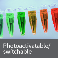
Photoconvertible fluorescent proteins

Dendra2 fluorescent protein vectors

Living Colors Dendra2 is a photoswitchable (green to red), monomeric fluorescent protein derived from octocoral Dendronephthya sp. The Dendra2 sequence has been optimized for maturation and a bright fluorescence both before and after photoswitching. Its codon usage is optimized for high expression in mammalian cells, but Dendra2 can be expressed in many other systems as well.

Living Colors Dendra2 is a photoswitchable (green to red), monomeric fluorescent protein derived from octocoral Dendronephthya sp. The Dendra2 sequence has been optimized for maturation and a bright fluorescence both before and after photoswitching. Its codon usage is optimized for high expression in mammalian cells, but Dendra2 can be expressed in many other systems as well.

Dendra2 matures efficiently both at 20 and 37°C, which makes it applicable to a wide range of experimental systems, from cultured mammalian cells to cold-blooded animals. Mammalian cells transiently transfected with Dendra2 display an evenly distributed green signal (e.g., without aggregation) within 10–12 hours post-transfection. No cell toxicity is observed.

Visualizing Dendra2 and Dendra2-tagged proteins

  • Visualizing green (unswitched) Dendra2
    Unconverted Dendra2 has excitation and emission maxima at 490 and 507 nm, respectively, similar to other green fluorescent proteins. Thus, an FITC (or analogous) filter set is ideal for visualizing green Dendra2.

    NOTE: Light of the same wavelength (intense blue light irradiation at 460–500 nm) is required to visualize and photoconvert Dendra2. Importantly, Dendra2 photoconversion occurs only at high light intensities, whereas Dendra2 green fluorescence can be detected at low light intensities. Be careful to select conditions that allow you to detect the green signal without undesirable photoconversion to red.
  • Switching Dendra2 from green to red
    You can photoconvert Dendra2 from green to red by light irradiation in either the UV (360–420 nm) or blue region (460–500 nm). We recommend that you use either a commonly available, 488-nm Ar laser line or a 405-nm diode laser (more efficient, but UV light can harm cells).
  • Visualizing red (switched) Dendra2
    Converted Dendra2 protein has excitation and emission maxima at 553 and 573 nm, respectively. Thus, a TRITC (or analogous) filter set can be used to visualize red Dendra2. Once it is irreversibly switched to its red form, Dendra2 is highly photostable. This makes it extremely useful for long-term protein tracking applications.

Excellent performance in fusion applications

Dendra2 is a monomeric fluorescent protein which has been successfully fused to a variety of proteins including cytoplasmic beta-actin, BH3 interacting domain death agonist (BID), nucleolar protein fibrillarin, vimentin, and alpha-tubulin.

High-contrast visualization

After complete photoconversion, the red fluorescence of Dendra2 increases 150–300 times, whereas the level of green fluorescence becomes 10–15 times lower. Thus, the increase in the red-to-green fluorescence ratio results in ~4,000-fold contrast. This provides a molecular tool to simultaneously track both the movement of the activated protein and its replacement with the non-activated form.

Dendra2 sequence files can be found in the Documents tab in the product table below.

 More  Less
Cat. # Product Size Price License Quantity Details
632546 pDendra2-C Vector 20 ug USD $596.00

pDendra2-C is a mammalian expression vector that encodes Dendra2, a green-to-red photoswitchable fluorescent protein derived from the octocoral Dendronephthya sp. An MCS at the 3' end of the coding sequence makes it possible to insert a gene of interest in order to express it as a C-terminal fusion with Dendra2. The unmodified vector can be used to express Dendra2 in mammalian cells.

Notice to purchaser

Our products are to be used for Research Use Only. They may not be used for any other purpose, including, but not limited to, use in humans, therapeutic or diagnostic use, or commercial use of any kind. Our products may not be transferred to third parties, resold, modified for resale, or used to manufacture commercial products or to provide a service to third parties without our prior written approval.

Documents Components Image Data

Back

Green-to-red photoconversion of Dendra2-tagged proteins

Green-to-red photoconversion of Dendra2-tagged proteins
Green-to-red photoconversion of Dendra2-tagged proteins. HeLa cells were transiently transfected with vectors encoding Dendra2-tagged fusion proteins. Dendra2 was converted to the red state in selected cells by brief illumination with a 488 nm laser. Confocal images were taken in the green and red channels after photoconversion.

Back

632546: pDendra2-C Vector

632546: pDendra2-C Vector

Back

Schematic for fluorescent fusion protein vectors

Schematic for fluorescent fusion protein vectors

Schematic for fluorescent fusion protein vectors. Panel A. N-terminal fluorescent protein (FP) vector. Panel B. C-terminal fluorescent protein vector.

632545 pDendra2-N Vector 20 ug USD $596.00

pDendra2-N is a mammalian expression vector that encodes Dendra2, a green-to-red photoswitchable fluorescent protein derived from the octocoral Dendronephthya sp. An MCS at the 5' end of the coding sequence makes it possible to insert a gene of interest in order to express it as an N-terminal fusion with Dendra2. The unmodified vector can be used to express Dendra2 in mammalian cells.

Notice to purchaser

Our products are to be used for Research Use Only. They may not be used for any other purpose, including, but not limited to, use in humans, therapeutic or diagnostic use, or commercial use of any kind. Our products may not be transferred to third parties, resold, modified for resale, or used to manufacture commercial products or to provide a service to third parties without our prior written approval.

Documents Components Image Data

Back

632545: pDendra2-N Vector

632545: pDendra2-N Vector

Back

Green-to-red photoconversion of Dendra2-tagged proteins

Green-to-red photoconversion of Dendra2-tagged proteins
Green-to-red photoconversion of Dendra2-tagged proteins. HeLa cells were transiently transfected with vectors encoding Dendra2-tagged fusion proteins. Dendra2 was converted to the red state in selected cells by brief illumination with a 488 nm laser. Confocal images were taken in the green and red channels after photoconversion.

Back

Schematic for fluorescent fusion protein vectors

Schematic for fluorescent fusion protein vectors

Schematic for fluorescent fusion protein vectors. Panel A. N-terminal fluorescent protein (FP) vector. Panel B. C-terminal fluorescent protein vector.

632544 pDendra2 Vector 20 ug USD $596.00

pDendra2 encodes Dendra2, a green-to-red photoswitchable fluorescent protein derived from the octocoral Dendronephthya sp. In this vector, the Dendra2 coding sequence can be excised using BamHI and HindIII, for transfer into another vector. The pDendra2 vector also contains a lac promoter sequence, and therefore can be used to express Dendra2 in E. coli. It is primarily intended to serve as a source of Dendra2 cDNA.

Notice to purchaser

Our products are to be used for Research Use Only. They may not be used for any other purpose, including, but not limited to, use in humans, therapeutic or diagnostic use, or commercial use of any kind. Our products may not be transferred to third parties, resold, modified for resale, or used to manufacture commercial products or to provide a service to third parties without our prior written approval.

Documents Components Image Data

Back

632544: pDendra2 Vector

632544: pDendra2 Vector

Back

Green-to-red photoconversion of Dendra2-tagged proteins

Green-to-red photoconversion of Dendra2-tagged proteins
Green-to-red photoconversion of Dendra2-tagged proteins. HeLa cells were transiently transfected with vectors encoding Dendra2-tagged fusion proteins. Dendra2 was converted to the red state in selected cells by brief illumination with a 488 nm laser. Confocal images were taken in the green and red channels after photoconversion.

Back

Basic fluorescent protein vector map

Basic fluorescent protein vector map

Basic fluorescent protein vector map. Use this bacterial expression vector as a source of the fluorescent protein gene. Note: There is a stop codon at the 5' end of the 3' MCS. The 3' MCS should not be used for cloning.

Selection guides Fluorescent protein vector finder
Selection guides Fluorescent protein quick guide

Overview

  • Living Colors Dendra2 is a green-to-red, high-contrast photoswitchable fluorescent protein
  • Irreversible switching from green to red is activated by UV or blue light
  • Switched (red) form is highly photostable
  • Matures at a wide range of temperatures
  • Monomeric fluorescent protein; suitable for fusions

More Information

Applications

  • Tracking cell, organelle, and protein movement
  • Visualizing real-time protein dynamics (movement, degradation, etc.)
  • Monitoring selective cell fate
  • Determining protein half-life

Additional product information

Please see the product's Certificate of Analysis for information about storage conditions, product components, and technical specifications. Please see the Kit Components List to determine kit components. Certificates of Analysis and Kit Components Lists are located under the Documents tab.


In-Fusion Cloning technology

Cloning shouldn't be work, it should just work ...

With In-Fusion technology, you will be able to perform directional, seamless cloning of any PCR fragment—or multiple fragments—into any linearized vector in a single 15-minute reaction, with >95% cloning efficiency. Our In-Fusion bundles are complete systems that include all of the reagents you need for your cloning experiments: PCR enzyme, competent cells, and a kit for PCR cleanup or gel extraction. We have optimized the reagents to work together to give you the right clone the first time.

In-Fusion Snap Assembly

Takara Bio USA, Inc.
United States/Canada: +1.800.662.2566 • Asia Pacific: +1.650.919.7300 • Europe: +33.(0)1.3904.6880 • Japan: +81.(0)77.565.6999
FOR RESEARCH USE ONLY. NOT FOR USE IN DIAGNOSTIC PROCEDURES. © 2025 Takara Bio Inc. All Rights Reserved. All trademarks are the property of Takara Bio Inc. or its affiliate(s) in the U.S. and/or other countries or their respective owners. Certain trademarks may not be registered in all jurisdictions. Additional product, intellectual property, and restricted use information is available at takarabio.com.

Takara Bio

Takara Bio USA, Inc. provides kits, reagents, instruments, and services that help researchers explore questions about gene discovery, regulation, and function. As a member of the Takara Bio Group, Takara Bio USA is part of a company that holds a leadership position in the global market and is committed to improving the human condition through biotechnology. Our mission is to develop high-quality innovative tools and services to accelerate discovery.

FOR RESEARCH USE ONLY. NOT FOR USE IN DIAGNOSTIC PROCEDURES (EXCEPT AS SPECIFICALLY NOTED).

Support
  • Contact us
  • Technical support
  • Customer service
  • Shipping & delivery
  • Sales
  • Feedback
Products
  • New products
  • Special offers
  • Instrument & reagent services
Learning centers
  • NGS
  • Gene function
  • Stem cell research
  • Protein research
  • PCR
  • Cloning
  • Nucleic acid purification
About
  • Our brands
  • Careers
  • Events
  • Blog
  • Need help?
  • Announcements
  • Quality and compliance
  • That's Good Science!
Facebook Twitter  LinkedIn

logo strip white

©2025 Takara Bio Inc. All Rights Reserved.

Region - North America Privacy Policy Terms and Conditions Terms of Use

Top


  • COVID-19 research
  • Viral detection with qPCR
  • SARS-CoV-2 pseudovirus
  • Human ACE2 stable cell line
  • Viral RNA isolation
  • Viral and host sequencing
  • Vaccine development
  • CRISPR screening
  • Drug discovery
  • Immune profiling
  • Publications
  • Next-generation sequencing
  • Spatial omics
  • RNA-seq
  • DNA-seq
  • Single-cell NGS automation
  • Reproductive health
  • Bioinformatics tools
  • Immune profiling
  • Real-time PCR
  • Great value master mixes
  • Signature enzymes
  • High-throughput real-time PCR solutions
  • Detection assays
  • References, standards, and buffers
  • Stem cell research
  • Media, differentiation kits, and matrices
  • Stem cells and stem cell-derived cells
  • mRNA and cDNA synthesis
  • In vitro transcription
  • cDNA synthesis kits
  • Reverse transcriptases
  • RACE kits
  • Purified cDNA & genomic DNA
  • Purified total RNA and mRNA
  • PCR
  • Most popular polymerases
  • High-yield PCR
  • High-fidelity PCR
  • GC rich PCR
  • PCR master mixes
  • Cloning
  • In-Fusion seamless cloning
  • Competent cells
  • Ligation kits
  • Restriction enzymes
  • Nucleic acid purification
  • Automated platforms
  • Plasmid purification kits
  • Genomic DNA purification kits
  • DNA cleanup kits
  • RNA purification kits
  • Gene function
  • Gene editing
  • Viral transduction
  • Fluorescent proteins
  • T-cell transduction and culture
  • Tet-inducible expression systems
  • Transfection reagents
  • Cell biology assays
  • Protein research
  • Purification products
  • Two-hybrid and one-hybrid systems
  • Mass spectrometry reagents
  • Antibodies and ELISAs
  • Primary antibodies and ELISAs by research area
  • Fluorescent protein antibodies
  • New products
  • Special offers
  • OEM
  • Portfolio
  • Process
  • Facilities
  • Request samples
  • FAQs
  • Instrument services
  • Apollo services
  • ICELL8 services
  • SmartChip ND system services
  • Gene and cell therapy manufacturing services
  • Services
  • Facilities
  • Our process
  • Resources
  • Customer service
  • Sales
  • Make an appointment with your sales rep
  • Shipping & delivery
  • Technical support
  • Feedback
  • Online tools
  • GoStix Plus FAQs
  • Partnering & Licensing
  • Vector information
  • Vector document overview
  • Vector document finder
Takara Bio's award-winning GMP-compliant manufacturing facility in Kusatsu, Shiga, Japan.

Partner with Takara Bio!

Takara Bio is proud to offer GMP-grade manufacturing capabilities at our award-winning facility in Kusatsu, Shiga, Japan.

  • Automation systems
  • Shasta Single Cell System introduction
  • SmartChip Real-Time PCR System introduction
  • ICELL8 introduction
  • Next-generation sequencing
  • RNA-seq
  • Technical notes
  • Technology and application overviews
  • FAQs and tips
  • DNA-seq protocols
  • Bioinformatics resources
  • Webinars
  • Spatial biology
  • Real-time PCR
  • Download qPCR resources
  • Overview
  • Reaction size guidelines
  • Guest webinar: extraction-free SARS-CoV-2 detection
  • Technical notes
  • Nucleic acid purification
  • Nucleic acid extraction webinars
  • Product demonstration videos
  • Product finder
  • Plasmid kit selection guide
  • RNA purification kit finder
  • mRNA and cDNA synthesis
  • mRNA synthesis
  • cDNA synthesis
  • PCR
  • Citations
  • PCR selection guide
  • Technical notes
  • FAQ
  • Cloning
  • Automated In-Fusion Cloning
  • In-Fusion Cloning general information
  • Primer design and other tools
  • In‑Fusion Cloning tips and FAQs
  • Applications and technical notes
  • Stem cell research
  • Overview
  • Protocols
  • Technical notes
  • Gene function
  • Gene editing
  • Viral transduction
  • T-cell transduction and culture
  • Inducible systems
  • Cell biology assays
  • Protein research
  • Capturem technology
  • Antibody immunoprecipitation
  • His-tag purification
  • Other tag purification
  • Expression systems
  • Antibodies and ELISA
  • Molecular diagnostics
  • Interview: adapting to change with Takara Bio
  • Applications
  • Solutions
  • Partnering
  • Contact us
  • mRNA and protein therapeutics
  • Characterizing the viral genome and host response
  • Identifying and cloning protein targets
  • Expressing and purifying protein targets
  • Immunizing mice and optimizing vaccines
  • Pathogen detection
  • Sample prep
  • Detection methods
  • Identification and characterization
  • SARS-CoV-2
  • Antibiotic-resistant bacteria
  • Food crop pathogens
  • Waterborne disease outbreaks
  • Viral-induced cancer
  • Immunotherapy research
  • T-cell therapy
  • Antibody therapeutics
  • T-cell receptor profiling
  • TBI initiatives in cancer therapy
  • Cancer research
  • Kickstart your cancer research with long-read sequencing
  • Sample prep from FFPE tissue
  • Sample prep from plasma
  • Cancer biomarker quantification
  • Single cancer cell analysis
  • Cancer transcriptome analysis
  • Cancer genomics and epigenomics
  • HLA typing in cancer
  • Gene editing for cancer therapy/drug discovery
  • Alzheimer's disease research
  • Antibody engineering
  • Sample prep from FFPE tissue
  • Single-cell sequencing
  • Reproductive health technologies
  • Embgenix FAQs
  • Preimplantation genetic testing
  • ESM partnership program
  • ESM Collection Kit forms
  • Infectious diseases
  • Develop vaccines for HIV
Create a web account with us

Log in to enjoy additional benefits

Want to save this information?

An account with takarabio.com entitles you to extra features such as:

•  Creating and saving shopping carts
•  Keeping a list of your products of interest
•  Saving all of your favorite pages on the site*
•  Accessing restricted content

*Save favorites by clicking the star () in the top right corner of each page while you're logged in.

Create an account to get started

  • BioView blog
  • Automation
  • Cancer research
  • Career spotlights
  • Current events
  • Customer stories
  • Gene editing
  • Research news
  • Single-cell analysis
  • Stem cell research
  • Tips and troubleshooting
  • Women in STEM
  • That's Good Support!
  • About our blog
  • That's Good Science!
  • SMART-Seq Pro Biomarker Discovery Contest
  • DNA extraction educational activity
  • That's Good Science Podcast
  • Season one
  • Season two
  • Season three
  • Our brands
  • Our history
  • In the news
  • Events
  • Biomarker discovery events
  • Calendar
  • Conferences
  • Speak with us
  • Careers
  • Company benefits
  • Trademarks
  • License statements
  • Quality statement
  • HQ-grade reagents
  • International Contacts by Region
  • United States and Canada
  • China
  • Japan
  • Korea
  • Europe
  • India
  • Affiliates & distributors
  • Need help?
  • Privacy request
  • Website FAQs

That's GOOD Science!

What does it take to generate good science? Careful planning, dedicated researchers, and the right tools. At Takara Bio, we thoughtfully develop exceptional products to tackle your most challenging research problems, and have an expert team of technical support professionals to help you along the way, all at superior value.

Explore what makes good science possible

 Customer Login
 View Cart (0)
Takara Bio
  • Home
  • Products
  • Services & Support
  • Learning centers
  • APPLICATIONS
  • About
  • Contact Us
  •  Customer Login
  • Register
  •  View Cart (0)

Takara Bio USA, Inc. provides kits, reagents, instruments, and services that help researchers explore questions about gene discovery, regulation, and function. As a member of the Takara Bio Group, Takara Bio USA is part of a company that holds a leadership position in the global market and is committed to improving the human condition through biotechnology. Our mission is to develop high-quality innovative tools and services to accelerate discovery.

FOR RESEARCH USE ONLY. NOT FOR USE IN DIAGNOSTIC PROCEDURES (EXCEPT AS SPECIFICALLY NOTED).

Clontech, TaKaRa, cellartis

  • Products
  • COVID-19 research
  • Next-generation sequencing
  • Real-time PCR
  • Stem cell research
  • mRNA and cDNA synthesis
  • PCR
  • Cloning
  • Nucleic acid purification
  • Gene function
  • Protein research
  • Antibodies and ELISA
  • New products
  • Special offers
  • COVID-19 research
  • Viral detection with qPCR
  • SARS-CoV-2 pseudovirus
  • Human ACE2 stable cell line
  • Viral RNA isolation
  • Viral and host sequencing
  • Vaccine development
  • CRISPR screening
  • Drug discovery
  • Immune profiling
  • Publications
  • Next-generation sequencing
  • Spatial omics
  • RNA-seq
  • DNA-seq
  • Single-cell NGS automation
  • Reproductive health
  • Bioinformatics tools
  • Immune profiling
  • Real-time PCR
  • Great value master mixes
  • Signature enzymes
  • High-throughput real-time PCR solutions
  • Detection assays
  • References, standards, and buffers
  • Stem cell research
  • Media, differentiation kits, and matrices
  • Stem cells and stem cell-derived cells
  • mRNA and cDNA synthesis
  • In vitro transcription
  • cDNA synthesis kits
  • Reverse transcriptases
  • RACE kits
  • Purified cDNA & genomic DNA
  • Purified total RNA and mRNA
  • PCR
  • Most popular polymerases
  • High-yield PCR
  • High-fidelity PCR
  • GC rich PCR
  • PCR master mixes
  • Cloning
  • In-Fusion seamless cloning
  • Competent cells
  • Ligation kits
  • Restriction enzymes
  • Nucleic acid purification
  • Automated platforms
  • Plasmid purification kits
  • Genomic DNA purification kits
  • DNA cleanup kits
  • RNA purification kits
  • Gene function
  • Gene editing
  • Viral transduction
  • Fluorescent proteins
  • T-cell transduction and culture
  • Tet-inducible expression systems
  • Transfection reagents
  • Cell biology assays
  • Protein research
  • Purification products
  • Two-hybrid and one-hybrid systems
  • Mass spectrometry reagents
  • Antibodies and ELISA
  • Primary antibodies and ELISAs by research area
  • Fluorescent protein antibodies
  • Services & Support
  • OEM
  • Instrument services
  • Gene and cell therapy manufacturing
  • Customer service
  • Sales
  • Shipping & delivery
  • Technical support
  • Feedback
  • Online tools
  • Partnering & Licensing
  • Vector information
  • OEM
  • Portfolio
  • Process
  • Facilities
  • Request samples
  • FAQs
  • Instrument services
  • Apollo services
  • ICELL8 services
  • SmartChip ND system services
  • Gene and cell therapy manufacturing
  • Services
  • Facilities
  • Our process
  • Resources
  • Sales
  • Make an appointment with your sales rep
  • Online tools
  • GoStix Plus FAQs
  • Vector information
  • Vector document overview
  • Vector document finder
  • Learning centers
  • Automation systems
  • Next-generation sequencing
  • Spatial biology
  • Real-time PCR
  • Nucleic acid purification
  • mRNA and cDNA synthesis
  • PCR
  • Cloning
  • Stem cell research
  • Gene function
  • Protein research
  • Antibodies and ELISA
  • Automation systems
  • Shasta Single Cell System introduction
  • SmartChip Real-Time PCR System introduction
  • ICELL8 introduction
  • Next-generation sequencing
  • RNA-seq
  • Technical notes
  • Technology and application overviews
  • FAQs and tips
  • DNA-seq protocols
  • Bioinformatics resources
  • Webinars
  • Real-time PCR
  • Download qPCR resources
  • Overview
  • Reaction size guidelines
  • Guest webinar: extraction-free SARS-CoV-2 detection
  • Technical notes
  • Nucleic acid purification
  • Nucleic acid extraction webinars
  • Product demonstration videos
  • Product finder
  • Plasmid kit selection guide
  • RNA purification kit finder
  • mRNA and cDNA synthesis
  • mRNA synthesis
  • cDNA synthesis
  • PCR
  • Citations
  • PCR selection guide
  • Technical notes
  • FAQ
  • Cloning
  • Automated In-Fusion Cloning
  • In-Fusion Cloning general information
  • Primer design and other tools
  • In‑Fusion Cloning tips and FAQs
  • Applications and technical notes
  • Stem cell research
  • Overview
  • Protocols
  • Technical notes
  • Gene function
  • Gene editing
  • Viral transduction
  • T-cell transduction and culture
  • Inducible systems
  • Cell biology assays
  • Protein research
  • Capturem technology
  • Antibody immunoprecipitation
  • His-tag purification
  • Other tag purification
  • Expression systems
  • APPLICATIONS
  • Molecular diagnostics
  • mRNA and protein therapeutics
  • Pathogen detection
  • Immunotherapy research
  • Cancer research
  • Alzheimer's disease research
  • Reproductive health technologies
  • Infectious diseases
  • Molecular diagnostics
  • Interview: adapting to change with Takara Bio
  • Applications
  • Solutions
  • Partnering
  • Contact us
  • mRNA and protein therapeutics
  • Characterizing the viral genome and host response
  • Identifying and cloning protein targets
  • Expressing and purifying protein targets
  • Immunizing mice and optimizing vaccines
  • Pathogen detection
  • Sample prep
  • Detection methods
  • Identification and characterization
  • SARS-CoV-2
  • Antibiotic-resistant bacteria
  • Food crop pathogens
  • Waterborne disease outbreaks
  • Viral-induced cancer
  • Immunotherapy research
  • T-cell therapy
  • Antibody therapeutics
  • T-cell receptor profiling
  • TBI initiatives in cancer therapy
  • Cancer research
  • Kickstart your cancer research with long-read sequencing
  • Sample prep from FFPE tissue
  • Sample prep from plasma
  • Cancer biomarker quantification
  • Single cancer cell analysis
  • Cancer transcriptome analysis
  • Cancer genomics and epigenomics
  • HLA typing in cancer
  • Gene editing for cancer therapy/drug discovery
  • Alzheimer's disease research
  • Antibody engineering
  • Sample prep from FFPE tissue
  • Single-cell sequencing
  • Reproductive health technologies
  • Embgenix FAQs
  • Preimplantation genetic testing
  • ESM partnership program
  • ESM Collection Kit forms
  • Infectious diseases
  • Develop vaccines for HIV
  • About
  • BioView blog
  • That's Good Science!
  • Our brands
  • Our history
  • In the news
  • Events
  • Careers
  • Trademarks
  • License statements
  • Quality and compliance
  • HQ-grade reagents
  • International Contacts by Region
  • Need help?
  • Website FAQs
  • BioView blog
  • Automation
  • Cancer research
  • Career spotlights
  • Current events
  • Customer stories
  • Gene editing
  • Research news
  • Single-cell analysis
  • Stem cell research
  • Tips and troubleshooting
  • Women in STEM
  • That's Good Support!
  • About our blog
  • That's Good Science!
  • SMART-Seq Pro Biomarker Discovery Contest
  • DNA extraction educational activity
  • That's Good Science Podcast
  • Season one
  • Season two
  • Season three
  • Events
  • Biomarker discovery events
  • Calendar
  • Conferences
  • Speak with us
  • Careers
  • Company benefits
  • International Contacts by Region
  • United States and Canada
  • China
  • Japan
  • Korea
  • Europe
  • India
  • Affiliates & distributors
  • Need help?
  • Privacy request
Takara Bio
  • Products
  • Services & Support
  • Learning centers
  • APPLICATIONS
  • About
  • Contact Us