Performance
Four vials of Cellartis cardiomyocytes were thawed and yielded 1.5 x 10⁷ viable cells. With 10% loss during pipetting, this accounted for 14 Cellartis EHTs. Within nine days, the spontaneously beating cells formed a syncytium and macroscopic contractions of the EHT was measured with the EHT analysis instrument (Figure 1).

Figure 1. Contraction analysis of Cellartis-derived engineered heart tissue (EHT). Left: live image of an EHT during contraction analysis. The EHT is spanning between two flexible silicone posts where optical figure recognition (blue crosses) is based and illuminated by an LED. Scale bar = 1 mm. Right: exemplary contraction peaks of a spontaneously beating EHT showing contraction force over time.
As known from other EHT studies (Hansen et al. 2010, Mannhardt et al. 2016), EHT contractions became stronger during culture. After three weeks in culture, the tissues were spontaneously beating with 43 ± 7 beats per minute, 0.15 ± 0.03 mN in contraction forces, a contraction time (T1) of 176 ± 5 ms, a relaxation time (T2) of 401 ± 42 ms, a contraction velocity of 1.3 ± 0.2 mN/sec, and a relaxation velocity of 0.7 ± 0.1 mN/sec (data are mean ± SD and n=13; Figure 2). The beating pattern was highly regular (low RR' index; Figure 2).

Figure 2. Baseline contractility data from Cellartis-cardiomyocyte-derived engineered heart tissue (EHT).
Characterization
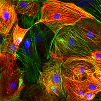
Whole mount immunofluorescence staining of Cellartis EHTs revealed elongation of the cells with good sarcomeric organization and distinct actinin and myosin light chain 2v (MLC2v) patterning (Figure 3).

Figure 3. Immunofluorescence characterization of Cellartis cardiomyocytes cultivated into EHT. Green: anti-α-actinin; red: anti-MLC2v; and blue: DRAQ5. Scale bar = 5 µm.