We use cookies to improve your browsing experience and provide meaningful content. Read our cookie policy. Accept
  •  Customer Login
  • Register
  •  View Cart (0)
  •  Customer Login
  • Register
  •  View Cart (0)

Takara Bio
  • Products
  • Services & Support
  • Learning centers
  • APPLICATIONS
  • About
  • Contact Us

Clontech Takara Cellartis

Close

  • ‹ Back to Sample preparation protocols
  • Basic cell preparation for the ICELL8 cx system
  • Protocol: Nuclei isolation from mammalian cells
  • Protocol: Mouse cardiomyocyte preparation
  • Isolate cells of any size
Single cardiomyocyte transcriptome Automating cardiomyocyte research
ICELL8 Innovators Series: scRNA-seq of cardiomyocytes Exploring cardiomyocyte transcriptomes with the ICELL8 system
ICELL8 system large-bore dispenser Blog: Keeping cardiac research pumping on the ICELL8 system
Identifying transcription factors involved in cardiomyocyte heterogeneity Blog: unlocking cardiomyocyte heterogeneity
Home › Learning centers › Automation systems › ICELL8 introduction › Sample preparation protocols › Protocol: Mouse cardiomyocyte preparation

ICELL8 introduction

  • ICELL8 cx applications
    • Archival nanowell sequencing
  • ICELL8 technology overview
  • ICELL8 cx technical specifications
  • ICELL8 technical specifications (original system)
  • ICELL8 system vs plate-seq
  • Webinars
    • Webinar: Leveraging single-cell transcriptomics and epigenomics for biomarker discovery
    • Advances in single-cell indexing registration
    • Single-Cell Workshop at 2020 NextGen Omics Series UK
    • The power of full-length scRNA-seq
    • Sign up: cardiomyocyte webinar
  • Technical notes
    • Enhancing biomarker discovery with SMART-Seq Pro kit and ICELL8 cx system
    • ICELL8 cx system target enrichment for fusions
    • ICELL8 cx system reagent formulation and dispense guidelines
    • Improved detection of gene fusions, SNPs, and alternative splicing
    • Full-length transcriptome analysis
    • High-throughput single-cell ATAC-seq
    • Protocol: High-throughput single-cell ATAC-Seq
    • Single-cell identification with CellSelect Software
    • Single-cell analysis elucidates cardiomyocyte differentiation from iPSCs
    • Combined TCR profiling and 5’ DE in single cells
    • Automated, high-throughput TCR profiling
  • Sample preparation protocols
    • Basic cell preparation for the ICELL8 cx system
    • Protocol: Nuclei isolation from mammalian cells
    • Protocol: Mouse cardiomyocyte preparation
    • Isolate cells of any size
  • Video resources
  • Citations
  • Posters
  • System & software notices
New products
Need help?
Contact Sales
Single cardiomyocyte transcriptome Automating cardiomyocyte research
ICELL8 Innovators Series: scRNA-seq of cardiomyocytes Exploring cardiomyocyte transcriptomes with the ICELL8 system
ICELL8 system large-bore dispenser Blog: Keeping cardiac research pumping on the ICELL8 system
Identifying transcription factors involved in cardiomyocyte heterogeneity Blog: unlocking cardiomyocyte heterogeneity
User-generated protocol

Protocol: Preparation of adult mouse cardiomyocytes for the ICELL8 system

Introduction Protocol Materials required References

Introduction  

The ICELL8 Single-Cell System is an open platform scientists are using to develop customized protocols, and we are very excited about the collaborations forming as a result of the system's flexibility. Takara Bio R&D scientists Kazu Tori and Montse Morell visited the Max Planck Institute for Heart and Lung Research in Bad Nauheim, Germany, to be trained by Dr. Stefan Günther of the Thomas Braun lab on a protocol they developed (Yekelchyk et al. 2019) for the preparation of cardiomyocytes for processing on the ICELL8 Single-Cell System. We are thankful for the team's hard work and willingness to share their workflow with us and other ICELL8 system users.

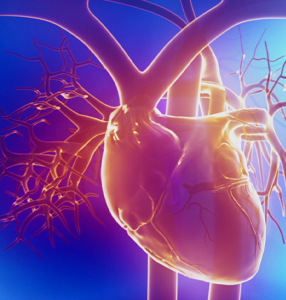
Primary adult mouse cardiomyocytes are a challenging cell type to capture, as they are delicate, sometimes multinucleated, and difficult to isolate as a pure population. They are also very large, and therefore a good candidate for isolation with the ICELL8 system dispenser's wide-bore nozzles. Furthermore, the ICELL8 system allows the user to visualize an isolated cardiomyocyte in a well and decide whether to process it. This protocol describes the isolation of cardiomyocytes from an adult mouse heart in preparation for dispensing and analysis on the ICELL8 system. The isolation protocol is mainly adapted from the published work by O'Connell, Rodrigo, and Simpson in 2007. If you wish to implement this protocol on the ICELL8 cx Single-Cell System, please contact your FSE.

Protocol  

A. Prior to isolation

The formulation of the Perfusion Buffer in this protocol differs slightly from the O'Connell protocol. (Please see the recipe, below.) The total volume of Perfusion Buffer to be prepared will be twice the capacity (in ml) of your chosen perfusion apparatus plus 10 ml. The following buffer is used during the whole process, but with different additives at various points.

  1. Prepare Perfusion Buffer as follows:

    113 mM NaCl
    4.7 mM KCl
    0.6 mM KH2PO4
    0.6 mM Na2HPO4
    1.2 mM MgSO4 x 7H2O
    12 mM NaHCO3
    10 mM KHCO3
    10 mM Hepes
    30 mM Taurine
    Add fresh before use:
    10 mM 2,3-Butanedione monoxime (BDM)*
    5.5 mM Glucose

    NOTE: BDM is used to decrease the contraction of the heart. The absence of Ca2+ is also important throughout the whole isolation process.


B. Collecting the sample

To aid the user in performing this subprotocol, here is a similar visual protocol (Judd, Lovas, and Huang 2016). Before beginning this subprotocol, prepare enough Perfusion Buffer for your perfusion apparatus. Also, prepare an equivalent volume of Digestion Buffer (for tissue digestion) by resuspending Liberase enzyme in Perfusion Buffer to a final concentration of 0.25 mg/ml and place on ice. Additionally, prepare 10 ml of Perfusion Buffer with 10% calf serum (for stopping digestion) and place on ice.

  1. Perform a peritoneal injection of heparin and anesthesia to the mouse. After the mouse is unconscious, extract the heart, ligate the right ventricle, and introduce a cannula in the aorta (so that the tip of the cannula is just above the aortic valve). Immediately start the perfusion with the Perfusion Buffer.
  2. Approximately 30 min after the start of perfusion, when the perfusion flow is faster, begin the digestion by exchanging the Digestion Buffer with the running Perfusion Buffer.
  3. Check the progress of digestion by looking at the flowthrough under the microscope for the presence of dissociated cells. The total digestion time will depend on the heart properties (mouse age, fibrotic stage, etc.) When rod-shaped single cells are present, and the heart looks pale, swollen, and flaccid, please proceed to the next step.
  4. At this point, remove the heart from the perfusion apparatus and place it in a watch glass containing 10 ml of Digestion Buffer.
  5. Carefully dissolve the heart by teasing it apart with tweezers and pipetting the buffer onto the tissue very slowly and gently with a 10-ml pipette. Cardiomyocytes are extremely delicate, so avoid vigorous pipetting.
  6. Transfer the solution to a 50-ml Falcon tube. Add the same volume of fresh Perfusion Buffer + serum (prepared earlier) to stop the digestion. The final concentration of serum should be 5%.
  7. After a few minutes, the majority of the cardiomyocytes will settle to the bottom. Carefully transfer the majority of the supernatant (containing additional cardiomyocytes) to a 15-ml tube and centrifuge at 500g for 1 min. This step is performed so as to recover as many cardiomyocytes as possible.
  8. Decant off the supernatant (contains smooth muscle cells and fibroblasts) and pour the obtained pellet into the 50-ml tube from Step 6.
  9. Filter the cell suspension with a 100-µM sieve.
  10. Count the cells using a Sedgewick-Rafter counting chamber or a standard hemocytometer using a 1:100 dilution of the cell suspension in buffer. Under the microscope, rod-shaped and round cells will be detected along with some cell debris. Only the rod-shaped cells should be considered healthy cardiomyocytes and, in a good sample, the percentage of rod-shaped cells should be higher than round cells. Write down the cell count here, as it may be useful later in Step C.5.a.

C. Staining and counting cells

  1. Perform the cell staining for a total of 5 min at room temperature. Add 1 µM CellTracker dye (1:1,000 of 1-mM stock) to label the cellular cytoplasm and Hoechst dye (1 drop per ml) to the cell suspension. If the cells start to sediment, invert the tube after dye addition; do not pipette.
  2. Wash the cells with Perfusion Buffer. Centrifuge the cells at 100g for 30 sec, and then pipette off the supernatant. (Alternatively, let the cardiomyocytes settle down for approximately 5 min at room temperature.) Add 1 ml of Perfusion Buffer then invert to resuspend (do not pipette). Repeat the wash twice with 1 ml of Perfusion Buffer for each wash.

    OPTIONAL: Check the staining under a fluorescence microscope, if desired. The cell cytoplasms should be red, and the nuclei should be blue.

  3. A final cell concentration is needed to calculate the dilution of cells for dispensing. Note the total volume of the cell suspension. Count the cells using one of the following options:

    1. Theoretical calculation: the survival rate throughout the isolation process tends to be about 40%, so the final cell number for dispensing can be calculated based on the following formula, which uses the initial cell count obtained after isolation in Step B.10.

      final cell concentration = (c * 0.375) (u / f)

      c = initial number of cells/ml, counted in Step B.10
      f = ml, final volume for resuspension of cells after washing
      u = ml, volume used for staining (e.g., 2 ml)

    2. Actual count: counting chamber or standard hemocytometer.

D. Dispensing cells

Please refer to operation and dispensing instructions in the ICELL8 system user manual.

  1. Dilute the cells before dispensing to achieve a lambda of 0.25 (in other words, 1 cell per 200 nl). To prepare 1 ml of this solution, use 0.4 U/µl RNase Inhibitor and 5X Second Diluent.

    NOTES:

    • The lambda used for cardiomyocytes is lower than usual for other cell types. Also, the concentration of diluent is higher than usual.
    • For negative and positive controls, use 0.4 U/µl RNase Inhibitor and 1X Second Diluent as in the user manual.
  2. Prepare the source plate by adding 35 μl of cell suspension to each of the 8 sample wells. Add fiducial mix and negative and positive controls to the source plate.
  3. Add a pause step during the sample dispensing process by selecting the proper option in the software. After the first dispense, the software will display a message alerting the user that the pause has been activated. At this time, remove the source plate from the dispensing platform and replace the cell suspension with 35 µl of fresh cell suspension in each of the 8 sample wells, and carefully put the source plate back onto the dispensing platform.

    NOTE: Pausing may work differently for the ICELL8 cx Single-Cell System. Please contact your FSE for support.

  4. Next, press [Start] to resume the protocol. The pause prompt will reappear one more time; after the pause, discard the cell suspension in all wells and add another 35 µl/well of fresh cell suspension. Try to minimize the wait time between pipetting and dispensing of cells.

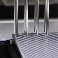
    IMPORTANT: This step is essential. Due to the size of cardiomyocytes, cells will quickly settle to the bottom; if the pause steps are not introduced, many cells will be aspirated in the first dispense, causing an unequal distribution (Figure 1).

    Figure 1. The image above depicts in gray the distribution of wells containing cells with and without the pause steps. Adding the pause steps results in significantly better cell capture.

  5. When all dispenses are complete, spin the plate at 300g for 3 min and proceed with imaging.

E. Imaging and manual triage

Refer to the manual triage instructions in the imaging software user manual.

  1. Adjust the channel settings for detection of CellTracker dye: set "gamma" to 0.8 and the "MinAutoContrastRange" to 255. The preset "DAPI" and "Texas Red" channels will not be used for this protocol. The exposure in the red channel can be adjusted for optimal visualization.
    Cardiomyocyte in ICELL8 well

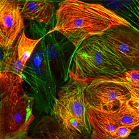
    Figure 2. Visualization of a rod-shaped, multinucleated cardiomyocyte in one well.


    NOTE: A typical ratio of binucleated to mononucleated cardiomyocytes is 80/20. The following criteria are suggested for manual triage: mono-, bi-, or multi-nucleated cells should have a rod shape; nuclei must overlap with the cytoplasm.


F. Preparing the library

Please refer to the ICELL8 3' DE Kit User Manual for detailed library prep instructions. For this protocol, use the following modifications:

  • During cell lysis, perform a preheat step by heating the chip at 72°C for 5 min, and then immediately place it on ice. This is important for optimal cell lysis.
  • Prepare the RT-PCR reaction with 2X concentration of Triton X-100.

Materials required  

  • Perfusion buffer ingredients, including 2,3-Butanedione monoxime (Sigma-Aldrich, Cat. # B0753)
  • Liberase T-Flex, Research Grade (Roche, Cat. # 05989132001)
  • Bovine calf serum (Sigma-Aldrich, Cat. # C8056-100ML)
  • EASYstrainer Cell Sieves (Greiner Bio-One, Cat # 542000)
  • Graticules S52 Sedgewick Rafter Counting Chamber (Glass Construction) (Spi Supplies, Cat. # 01010-AB)
  • CellTracker Red CMTPX Dye (Thermo Fisher Scientific, Cat. # C34552)

References  

Judd, J., Lovas, J. & Huang, G. N. Isolation, Culture and Transduction of Adult Mouse Cardiomyocytes. J. Vis. Exp. (2016). Watch the visual protocol »

O'Connell, T. D., Rodrigo, M. C. & Simpson, P. C. in Cardiovasc. Proteomics 271–296 (Humana Press, 2007).

Yekelchyk, M., Guenther, S., Preussner, J. & Braun, T. Mono- and multi-nucleated ventricular cardiomyocytes constitute a transcriptionally homogenous cell population. Basic Res. Cardiol. 114, 36 (2019). Read the paper »


User-generated protocols

User-generated protocols

User-generated protocols are based on internal proof-of-concept experiments, customer collaborations, and published literature. In some cases, relevant results are discussed in our research news BioView blog articles. While we expect these protocols to be successful in your hands, they may not be fully reviewed or optimized. We encourage you to contact us or refer to the published literature for more information about these user-generated and -reported protocols. 

If you are looking for a product-specific, fully optimized User Manual or Protocol-At-A-Glance, please visit the product's product page, open the item's product details row in the price table, and click Documents. More detailed instructions for locating documents are available on our website FAQs page.

Questions? Protocols of your own that you would like to share?

Contact technical support Give feedback

Takara Bio USA, Inc.
United States/Canada: +1.800.662.2566 • Asia Pacific: +1.650.919.7300 • Europe: +33.(0)1.3904.6880 • Japan: +81.(0)77.565.6999
FOR RESEARCH USE ONLY. NOT FOR USE IN DIAGNOSTIC PROCEDURES. © 2025 Takara Bio Inc. All Rights Reserved. All trademarks are the property of Takara Bio Inc. or its affiliate(s) in the U.S. and/or other countries or their respective owners. Certain trademarks may not be registered in all jurisdictions. Additional product, intellectual property, and restricted use information is available at takarabio.com.

Takara Bio

Takara Bio USA, Inc. provides kits, reagents, instruments, and services that help researchers explore questions about gene discovery, regulation, and function. As a member of the Takara Bio Group, Takara Bio USA is part of a company that holds a leadership position in the global market and is committed to improving the human condition through biotechnology. Our mission is to develop high-quality innovative tools and services to accelerate discovery.

FOR RESEARCH USE ONLY. NOT FOR USE IN DIAGNOSTIC PROCEDURES (EXCEPT AS SPECIFICALLY NOTED).

Support
  • Contact us
  • Technical support
  • Customer service
  • Shipping & delivery
  • Sales
  • Feedback
Products
  • New products
  • Special offers
  • Instrument & reagent services
Learning centers
  • NGS
  • Gene function
  • Stem cell research
  • Protein research
  • PCR
  • Cloning
  • Nucleic acid purification
About
  • Our brands
  • Careers
  • Events
  • Blog
  • Need help?
  • Announcements
  • Quality and compliance
  • That's Good Science!
Facebook Twitter  LinkedIn

logo strip white

©2025 Takara Bio Inc. All Rights Reserved.

Region - North America Privacy Policy Terms and Conditions Terms of Use

Top


  • COVID-19 research
  • Viral detection with qPCR
  • SARS-CoV-2 pseudovirus
  • Human ACE2 stable cell line
  • Viral RNA isolation
  • Viral and host sequencing
  • Vaccine development
  • CRISPR screening
  • Drug discovery
  • Immune profiling
  • Publications
  • Next-generation sequencing
  • Spatial omics
  • RNA-seq
  • DNA-seq
  • Single-cell NGS automation
  • Reproductive health
  • Bioinformatics tools
  • Immune profiling
  • Real-time PCR
  • Great value master mixes
  • Signature enzymes
  • High-throughput real-time PCR solutions
  • Detection assays
  • References, standards, and buffers
  • Stem cell research
  • Media, differentiation kits, and matrices
  • Stem cells and stem cell-derived cells
  • mRNA and cDNA synthesis
  • In vitro transcription
  • cDNA synthesis kits
  • Reverse transcriptases
  • RACE kits
  • Purified cDNA & genomic DNA
  • Purified total RNA and mRNA
  • PCR
  • Most popular polymerases
  • High-yield PCR
  • High-fidelity PCR
  • GC rich PCR
  • PCR master mixes
  • Cloning
  • In-Fusion seamless cloning
  • Competent cells
  • Ligation kits
  • Restriction enzymes
  • Nucleic acid purification
  • Automated platforms
  • Plasmid purification kits
  • Genomic DNA purification kits
  • DNA cleanup kits
  • RNA purification kits
  • Gene function
  • Gene editing
  • Viral transduction
  • Fluorescent proteins
  • T-cell transduction and culture
  • Tet-inducible expression systems
  • Transfection reagents
  • Cell biology assays
  • Protein research
  • Purification products
  • Two-hybrid and one-hybrid systems
  • Mass spectrometry reagents
  • Antibodies and ELISAs
  • Primary antibodies and ELISAs by research area
  • Fluorescent protein antibodies
  • New products
  • Special offers
  • OEM
  • Portfolio
  • Process
  • Facilities
  • Request samples
  • FAQs
  • Instrument services
  • Apollo services
  • ICELL8 services
  • SmartChip ND system services
  • Gene and cell therapy manufacturing services
  • Services
  • Facilities
  • Our process
  • Resources
  • Customer service
  • Sales
  • Make an appointment with your sales rep
  • Shipping & delivery
  • Technical support
  • Feedback
  • Online tools
  • GoStix Plus FAQs
  • Partnering & Licensing
  • Vector information
  • Vector document overview
  • Vector document finder
Takara Bio's award-winning GMP-compliant manufacturing facility in Kusatsu, Shiga, Japan.

Partner with Takara Bio!

Takara Bio is proud to offer GMP-grade manufacturing capabilities at our award-winning facility in Kusatsu, Shiga, Japan.

  • Automation systems
  • Shasta Single Cell System introduction
  • SmartChip Real-Time PCR System introduction
  • ICELL8 introduction
  • Next-generation sequencing
  • RNA-seq
  • Technical notes
  • Technology and application overviews
  • FAQs and tips
  • DNA-seq protocols
  • Bioinformatics resources
  • Webinars
  • Spatial biology
  • Real-time PCR
  • Download qPCR resources
  • Overview
  • Reaction size guidelines
  • Guest webinar: extraction-free SARS-CoV-2 detection
  • Technical notes
  • Nucleic acid purification
  • Nucleic acid extraction webinars
  • Product demonstration videos
  • Product finder
  • Plasmid kit selection guide
  • RNA purification kit finder
  • mRNA and cDNA synthesis
  • mRNA synthesis
  • cDNA synthesis
  • PCR
  • Citations
  • PCR selection guide
  • Technical notes
  • FAQ
  • Cloning
  • Automated In-Fusion Cloning
  • In-Fusion Cloning general information
  • Primer design and other tools
  • In‑Fusion Cloning tips and FAQs
  • Applications and technical notes
  • Stem cell research
  • Overview
  • Protocols
  • Technical notes
  • Gene function
  • Gene editing
  • Viral transduction
  • T-cell transduction and culture
  • Inducible systems
  • Cell biology assays
  • Protein research
  • Capturem technology
  • Antibody immunoprecipitation
  • His-tag purification
  • Other tag purification
  • Expression systems
  • Antibodies and ELISA
  • Molecular diagnostics
  • Interview: adapting to change with Takara Bio
  • Applications
  • Solutions
  • Partnering
  • Contact us
  • mRNA and protein therapeutics
  • Characterizing the viral genome and host response
  • Identifying and cloning protein targets
  • Expressing and purifying protein targets
  • Immunizing mice and optimizing vaccines
  • Pathogen detection
  • Sample prep
  • Detection methods
  • Identification and characterization
  • SARS-CoV-2
  • Antibiotic-resistant bacteria
  • Food crop pathogens
  • Waterborne disease outbreaks
  • Viral-induced cancer
  • Immunotherapy research
  • T-cell therapy
  • Antibody therapeutics
  • T-cell receptor profiling
  • TBI initiatives in cancer therapy
  • Cancer research
  • Kickstart your cancer research with long-read sequencing
  • Sample prep from FFPE tissue
  • Sample prep from plasma
  • Cancer biomarker quantification
  • Single cancer cell analysis
  • Cancer transcriptome analysis
  • Cancer genomics and epigenomics
  • HLA typing in cancer
  • Gene editing for cancer therapy/drug discovery
  • Alzheimer's disease research
  • Antibody engineering
  • Sample prep from FFPE tissue
  • Single-cell sequencing
  • Reproductive health technologies
  • Embgenix FAQs
  • Preimplantation genetic testing
  • ESM partnership program
  • ESM Collection Kit forms
  • Infectious diseases
  • Develop vaccines for HIV
Create a web account with us

Log in to enjoy additional benefits

Want to save this information?

An account with takarabio.com entitles you to extra features such as:

•  Creating and saving shopping carts
•  Keeping a list of your products of interest
•  Saving all of your favorite pages on the site*
•  Accessing restricted content

*Save favorites by clicking the star () in the top right corner of each page while you're logged in.

Create an account to get started

  • BioView blog
  • Automation
  • Cancer research
  • Career spotlights
  • Current events
  • Customer stories
  • Gene editing
  • Research news
  • Single-cell analysis
  • Stem cell research
  • Tips and troubleshooting
  • Women in STEM
  • That's Good Support!
  • About our blog
  • That's Good Science!
  • SMART-Seq Pro Biomarker Discovery Contest
  • DNA extraction educational activity
  • That's Good Science Podcast
  • Season one
  • Season two
  • Season three
  • Our brands
  • Our history
  • In the news
  • Events
  • Biomarker discovery events
  • Calendar
  • Conferences
  • Speak with us
  • Careers
  • Company benefits
  • Trademarks
  • License statements
  • Quality statement
  • HQ-grade reagents
  • International Contacts by Region
  • United States and Canada
  • China
  • Japan
  • Korea
  • Europe
  • India
  • Affiliates & distributors
  • Need help?
  • Privacy request
  • Website FAQs

That's GOOD Science!

What does it take to generate good science? Careful planning, dedicated researchers, and the right tools. At Takara Bio, we thoughtfully develop exceptional products to tackle your most challenging research problems, and have an expert team of technical support professionals to help you along the way, all at superior value.

Explore what makes good science possible

 Customer Login
 View Cart (0)
Takara Bio
  • Home
  • Products
  • Services & Support
  • Learning centers
  • APPLICATIONS
  • About
  • Contact Us
  •  Customer Login
  • Register
  •  View Cart (0)

Takara Bio USA, Inc. provides kits, reagents, instruments, and services that help researchers explore questions about gene discovery, regulation, and function. As a member of the Takara Bio Group, Takara Bio USA is part of a company that holds a leadership position in the global market and is committed to improving the human condition through biotechnology. Our mission is to develop high-quality innovative tools and services to accelerate discovery.

FOR RESEARCH USE ONLY. NOT FOR USE IN DIAGNOSTIC PROCEDURES (EXCEPT AS SPECIFICALLY NOTED).

Clontech, TaKaRa, cellartis

  • Products
  • COVID-19 research
  • Next-generation sequencing
  • Real-time PCR
  • Stem cell research
  • mRNA and cDNA synthesis
  • PCR
  • Cloning
  • Nucleic acid purification
  • Gene function
  • Protein research
  • Antibodies and ELISA
  • New products
  • Special offers
  • COVID-19 research
  • Viral detection with qPCR
  • SARS-CoV-2 pseudovirus
  • Human ACE2 stable cell line
  • Viral RNA isolation
  • Viral and host sequencing
  • Vaccine development
  • CRISPR screening
  • Drug discovery
  • Immune profiling
  • Publications
  • Next-generation sequencing
  • Spatial omics
  • RNA-seq
  • DNA-seq
  • Single-cell NGS automation
  • Reproductive health
  • Bioinformatics tools
  • Immune profiling
  • Real-time PCR
  • Great value master mixes
  • Signature enzymes
  • High-throughput real-time PCR solutions
  • Detection assays
  • References, standards, and buffers
  • Stem cell research
  • Media, differentiation kits, and matrices
  • Stem cells and stem cell-derived cells
  • mRNA and cDNA synthesis
  • In vitro transcription
  • cDNA synthesis kits
  • Reverse transcriptases
  • RACE kits
  • Purified cDNA & genomic DNA
  • Purified total RNA and mRNA
  • PCR
  • Most popular polymerases
  • High-yield PCR
  • High-fidelity PCR
  • GC rich PCR
  • PCR master mixes
  • Cloning
  • In-Fusion seamless cloning
  • Competent cells
  • Ligation kits
  • Restriction enzymes
  • Nucleic acid purification
  • Automated platforms
  • Plasmid purification kits
  • Genomic DNA purification kits
  • DNA cleanup kits
  • RNA purification kits
  • Gene function
  • Gene editing
  • Viral transduction
  • Fluorescent proteins
  • T-cell transduction and culture
  • Tet-inducible expression systems
  • Transfection reagents
  • Cell biology assays
  • Protein research
  • Purification products
  • Two-hybrid and one-hybrid systems
  • Mass spectrometry reagents
  • Antibodies and ELISA
  • Primary antibodies and ELISAs by research area
  • Fluorescent protein antibodies
  • Services & Support
  • OEM
  • Instrument services
  • Gene and cell therapy manufacturing
  • Customer service
  • Sales
  • Shipping & delivery
  • Technical support
  • Feedback
  • Online tools
  • Partnering & Licensing
  • Vector information
  • OEM
  • Portfolio
  • Process
  • Facilities
  • Request samples
  • FAQs
  • Instrument services
  • Apollo services
  • ICELL8 services
  • SmartChip ND system services
  • Gene and cell therapy manufacturing
  • Services
  • Facilities
  • Our process
  • Resources
  • Sales
  • Make an appointment with your sales rep
  • Online tools
  • GoStix Plus FAQs
  • Vector information
  • Vector document overview
  • Vector document finder
  • Learning centers
  • Automation systems
  • Next-generation sequencing
  • Spatial biology
  • Real-time PCR
  • Nucleic acid purification
  • mRNA and cDNA synthesis
  • PCR
  • Cloning
  • Stem cell research
  • Gene function
  • Protein research
  • Antibodies and ELISA
  • Automation systems
  • Shasta Single Cell System introduction
  • SmartChip Real-Time PCR System introduction
  • ICELL8 introduction
  • Next-generation sequencing
  • RNA-seq
  • Technical notes
  • Technology and application overviews
  • FAQs and tips
  • DNA-seq protocols
  • Bioinformatics resources
  • Webinars
  • Real-time PCR
  • Download qPCR resources
  • Overview
  • Reaction size guidelines
  • Guest webinar: extraction-free SARS-CoV-2 detection
  • Technical notes
  • Nucleic acid purification
  • Nucleic acid extraction webinars
  • Product demonstration videos
  • Product finder
  • Plasmid kit selection guide
  • RNA purification kit finder
  • mRNA and cDNA synthesis
  • mRNA synthesis
  • cDNA synthesis
  • PCR
  • Citations
  • PCR selection guide
  • Technical notes
  • FAQ
  • Cloning
  • Automated In-Fusion Cloning
  • In-Fusion Cloning general information
  • Primer design and other tools
  • In‑Fusion Cloning tips and FAQs
  • Applications and technical notes
  • Stem cell research
  • Overview
  • Protocols
  • Technical notes
  • Gene function
  • Gene editing
  • Viral transduction
  • T-cell transduction and culture
  • Inducible systems
  • Cell biology assays
  • Protein research
  • Capturem technology
  • Antibody immunoprecipitation
  • His-tag purification
  • Other tag purification
  • Expression systems
  • APPLICATIONS
  • Molecular diagnostics
  • mRNA and protein therapeutics
  • Pathogen detection
  • Immunotherapy research
  • Cancer research
  • Alzheimer's disease research
  • Reproductive health technologies
  • Infectious diseases
  • Molecular diagnostics
  • Interview: adapting to change with Takara Bio
  • Applications
  • Solutions
  • Partnering
  • Contact us
  • mRNA and protein therapeutics
  • Characterizing the viral genome and host response
  • Identifying and cloning protein targets
  • Expressing and purifying protein targets
  • Immunizing mice and optimizing vaccines
  • Pathogen detection
  • Sample prep
  • Detection methods
  • Identification and characterization
  • SARS-CoV-2
  • Antibiotic-resistant bacteria
  • Food crop pathogens
  • Waterborne disease outbreaks
  • Viral-induced cancer
  • Immunotherapy research
  • T-cell therapy
  • Antibody therapeutics
  • T-cell receptor profiling
  • TBI initiatives in cancer therapy
  • Cancer research
  • Kickstart your cancer research with long-read sequencing
  • Sample prep from FFPE tissue
  • Sample prep from plasma
  • Cancer biomarker quantification
  • Single cancer cell analysis
  • Cancer transcriptome analysis
  • Cancer genomics and epigenomics
  • HLA typing in cancer
  • Gene editing for cancer therapy/drug discovery
  • Alzheimer's disease research
  • Antibody engineering
  • Sample prep from FFPE tissue
  • Single-cell sequencing
  • Reproductive health technologies
  • Embgenix FAQs
  • Preimplantation genetic testing
  • ESM partnership program
  • ESM Collection Kit forms
  • Infectious diseases
  • Develop vaccines for HIV
  • About
  • BioView blog
  • That's Good Science!
  • Our brands
  • Our history
  • In the news
  • Events
  • Careers
  • Trademarks
  • License statements
  • Quality and compliance
  • HQ-grade reagents
  • International Contacts by Region
  • Need help?
  • Website FAQs
  • BioView blog
  • Automation
  • Cancer research
  • Career spotlights
  • Current events
  • Customer stories
  • Gene editing
  • Research news
  • Single-cell analysis
  • Stem cell research
  • Tips and troubleshooting
  • Women in STEM
  • That's Good Support!
  • About our blog
  • That's Good Science!
  • SMART-Seq Pro Biomarker Discovery Contest
  • DNA extraction educational activity
  • That's Good Science Podcast
  • Season one
  • Season two
  • Season three
  • Events
  • Biomarker discovery events
  • Calendar
  • Conferences
  • Speak with us
  • Careers
  • Company benefits
  • International Contacts by Region
  • United States and Canada
  • China
  • Japan
  • Korea
  • Europe
  • India
  • Affiliates & distributors
  • Need help?
  • Privacy request
Takara Bio
  • Products
  • Services & Support
  • Learning centers
  • APPLICATIONS
  • About
  • Contact Us