We use cookies to improve your browsing experience and provide meaningful content. Read our cookie policy. Accept
  •  Customer Login
  • Register
  •  View Cart (0)
  •  Customer Login
  • Register
  •  View Cart (0)

Takara Bio
  • Products
  • Services & Support
  • Learning centers
  • APPLICATIONS
  • About
  • Contact Us

Clontech Takara Cellartis

Close

  • ‹ Back to Pluripotent stem cells
  • Using the DEF-CS system to culture human iPS cells
  • Comparison of the Cellartis DEF-CS system with other vendors' human iPS cell culture systems
  • Reprogramming PBMCs
  • Reprogramming fibroblasts
Technical notes Tech note: reprogramming fibroblasts
Protocols Protocol: reprogramming PBMCs
Protocols Protocol: reprogramming fibroblasts
Home › Learning centers › Stem cell research › Technical notes › Pluripotent stem cells › Reprogramming PBMCs

Technical notes

  • Pluripotent stem cells
    • Using the DEF-CS system to culture human iPS cells
    • Comparison of the Cellartis DEF-CS system with other vendors' human iPS cell culture systems
    • Reprogramming PBMCs
    • Reprogramming fibroblasts
  • Gene editing in hiPS cells
    • Tagging an endogenous gene with AcGFP1 in hiPS cells
    • Tagging an endogenous gene with a myc tag in hiPS cells
    • Generating clonal hiPS cell lines deficient in CD81
    • Introducing a tyrosinemia-related SNP in hiPS cells
    • Inserting an expression cassette into the AAVS1 locus in hiPS cells
    • Editing hiPS cells using electroporation
    • Editing hiPS cells using gesicle technology
    • Single-cell cloning of hiPS cells
  • Organoids
    • Retinal organoid differentiation from iPSCs cultured in the Cellartis DEF-CS 500 Culture System
    • Liver organoid differentiation from iPSCs for prediction of drug-induced liver injury
    • Generation of embryonic organoids using NDiff 227 neural differentiation medium
  • Beta cells
    • Beta cells for disease modeling
  • Hepatocytes
    • hiPS-HEP cells for disease modeling
    • hiPS-HEP cells for drug metabolism studies
    • Power medium for long-term human primary hepatocyte culture
    • iPS cell to hepatocyte differentiation system
  • Cardiomyocytes
    • Making engineered heart tissue with cardiomyocytes
  • Neural stem cells
    • RHB-A neural stem cell medium
New products
Need help?
Contact Sales
Technical notes Tech note: reprogramming fibroblasts
Protocols Protocol: reprogramming PBMCs
Protocols Protocol: reprogramming fibroblasts
Tech Note

Reprogramming of human PBMCs using Sendai virus and the Cellartis DEF-CS 500 Culture System

  • High success rate
    Reprogramming PBMCs using Sendai virus and the Cellartis DEF-CS 500 Culture System reliably generates multiple iPS cell colonies.
  • Robust expansion of clones
    The Cellartis DEF-CS 500 Culture System promotes a high survival rate of iPS cell colonies and robust proliferation of iPS cell clones.
  • Highly pluripotent iPS cells with robust differentiation capability
    The reprogrammed iPS cells show high expression levels of the stem cell markers Oct4, SSEA-4, TRA-1-60, and TRA-1-81.
  • Generated iPS cells also display expected differentiation patterns with the Cellartis Definitive Endoderm Differentiation Kit with DEF-CS Culture System and Cellartis Hepatocyte Differentiation Kit.
Introduction Results Conclusion Methods References

Introduction  

Induced pluripotent stem (iPS) cells are generated by reprogramming a differentiated somatic cell into a pluripotent stem cell. This source of pluripotent cells enables drug discovery, disease modeling, and ultimately gives us a chance to find cures for today’s most devastating diseases. With iPS cells, researchers can generate patient- or disease-specific cells, enabling new frontiers in disease modeling (Meissner, Wernig, and Jaenisch 2007).

Although there are several methods for reprogramming cells, the Sendai-virus method was shown to be highly reliable and efficient in comparison to other non-integrating methods (Schlaeger et al. 2015). The most commonly used reprogramming factors are the Yamanaka factors, Oct3/4, Sox2, c-Myc, and Klf4, which are suitable for efficient reprogramming (Takahashi et al. 2007).

The Cellartis DEF-CS 500 Culture System promotes reliable growth of hiPS cells in a defined, feeder-free environment. Cells are grown as a homogeneous monolayer and are enzymatically passaged as single cells that maintain pluripotency with a stable karyotype for more than 20 passages (Asplund et al. 2016). Here we show that the Cellartis DEF-CS 500 Culture System efficiently and robustly reprograms human primary peripheral blood mononuclear cells (PBMCs) using Sendai virus in a 21-day protocol (Figure 1).


Figure 1. Reprogramming timeline with the Cellartis DEF-CS 500 Culture System.

Results  

Using our PBMC reprogramming protocol (Figure 1), we reprogrammed seven different human PBMC cultures using Sendai virus and the Cellartis DEF-CS 500 Culture System. First, human PBMCs from ATCC were reprogrammed to evaluate the method, then PBMCs from 6 different donors (A–F) were reprogrammed. Most of the data shown are from A–F, which were analyzed in greater detail.

For all tested PBMCs, stable iPS cell lines were generated on the first transduction attempt. We transduced on the order of 106 PBMC cells from each donor, which generated large numbers of colonies, and picked 8–30 colonies from each of the donors for further culturing.

The survival rate of picked colonies from Donors A–F varies between 40–75%, depending on the donor, according to Table I. Here, the survival rate reflects the number of healthy, pluripotent colonies with robust growth.

Table I. The survival rate of picked colonies from donors A–F.
Survival rate of reprogrammed colonies
Donor A B C D E F
Picked colonies 15 12 12 12 8 8
Generated iPS cell lines 6 9 8 5 6 6
Generated iPS cell lines (%) 40 75 67 41 75 75

The morphology of the cells was monitored regularly. Figure 2 shows the morphological changes of a colony from the day of picking until the clone is ready to be frozen as a seed bank at Passage 6.

Morphological changes of a colony from picking to freezing

Figure 2. The progress from a colony at the day of picking to a clone ready to be frozen as a seed bank.

One generated iPS cell line from each donor—six iPS cell lines in total—were further passaged to study the loss of the Sendai virus. At some passages the amount of Sendai virus RNA was measured in the cells using quantitative real-time PCR. Ct values ≥36 were considered negative. At Passage 7–8, two-thirds of the iPS cell lines were cleared from Sendai virus RNA. All tested iPS cell lines were cleared from Sendai virus RNA by Passage 18 (see Figure 3).

Sendai virus is undetectable after Passage 18

Figure 3. Sendai virus is undetectable after Passage 18. Sendai virus RNA in the iPS cell lines was continuously measured using quantitative real-time PCR. Ct values ≥36 were considered negative.

At Passage 15, the iPS cell lines were analyzed for the stem cell markers Oct4, SSEA-4, TRA-1-60, and TRA-1-81, using immunocytochemistry (ICC). A vast majority of the cells were positive for all four markers (see Figure 4), whereas no expression of SSEA-1 was observed.

Immunocytochemical detection of stem cell markers in an iPS cell line

Figure 4. Immunocytochemical analysis for the detection of the stem cell markers Oct4, SSEA-4, TRA-1-60 and TRA-1-81 in one of the iPS cell lines at Passage 15. SSEA-1 was used as a negative marker. DAPI was used for nuclear staining. Overlap of DAPI and markers is shown in purple.

At Passages 14–17, the six iPS cell lines were also analyzed for the expression of Oct4 by flow cytometry. The cells contained a high proportion of Oct4-positive cells, according to Figure 5.

Analysis of 6 iPS cell lines for Oct4 expression by flow cytometry

Figure 5. The 6 iPS cell lines were analyzed for the expression of Oct4 by flow cytometry. All cell lines were nearly 100% positive for Oct4. Panel A. Representative FACS plot. Panel B. The percentage of cells positive for Oct4 varied in a narrow range between 99.58% and 99.94%.

To further examine the pluripotency of the six generated iPS cell lines, they were differentiated into Definitive Endoderm (DE) cells and then further into hepatocytes. All iPS cells generated DE cells and hepatocytes. ICC was performed to confirm differentiation to DE using the DE marker SOX17 and pluripotency marker Oct4. The majority of the cells in each iPS line were differentiated to DE cells, as indicated by positive staining for SOX17 and negative for Oct4 at Day 7, shown in Figure 6.

Differentiation to DE shown by morphology and Sox17, Oct4 markers

Figure 6. Differentiation to DE indicated by morphology, the DE marker Sox17 (red), and the pluripotency marker Oct4 (green). The majority of cells from each iPSC line was differentiated to DE cells. DAPI was used for nuclear staining.

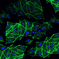
ICC was also performed to confirm differentiation to hepatocytes after 30 days of differentiation, as indicated by the hepatic markers HNF4α, albumin, and CYP3A4. The majority of the cells stained positive for HNF4α. A subset of the hepatocytes was also positive for albumin or CYP3A4, with only a few double-stained cells (see Figure 7).

Differentiation to hepatocytes shown by morphology and hepatic markers

Figure 7. Differentiation to hepatocytes after 30 days of differentiation indicated by morphology, the hepatic markers HNF4α, albumin (green), and CYP3A4 (red). The majority of the cells stained positive for HNF4α. A subset of the hepatocytes was also positive for albumin or CYP3A4, with only a few double-stained cells. DAPI was used for nuclear staining.

Conclusion  

Using the Cellartis DEF-CS 500 Culture System when reprogramming human PBMCs with Sendai virus allowed for generation of multiple iPS cell lines from each donor with only one transduction step. The Cellartis DEF-CS 500 Culture System promoted high rates of survival in the emerging iPS cell colonies and robust proliferation into iPS cell lines. The reprogrammed iPS cell lines showed high expression levels of the stem cell markers Oct4, SSEA-4, TRA-1-60, and TRA-1-81, and the typical morphology of undifferentiated stem cells. In addition, the generated iPS cells were shown to differentiate into definitive endoderm and hepatocytes when exposed to the Cellartis Definitive Endoderm Differentiation Kit with DEF-CS Culture System and the Cellartis Hepatocyte Differentiation Kit. The highly efficient and reliable performance of the Cellartis DEF-CS 500 Culture System in these studies demonstrates the time- and cost-effectiveness of this method.

Methods  

Culture of PBMCs

The PBMCs were thawed and seeded in PBMC medium. They were maintained in PBMC medium for four days prior to the start of the Sendai reprogramming.

Sendai reprogramming and transfer to the Cellartis DEF-CS 500 Culture System

The CytoTune-iPS 2.0 Sendai Reprogramming Kit (Fisher Scientific) was used to reprogram the PBMCs. The reprogramming vectors include the four Yamanaka factors, Oct3/4, Sox2, c-Myc, and Klf4. The major reprogramming steps are shown in Figure 1. Plating of the PBMCs, transduction, and maintenance of transduced cells until Day 3 were performed according to the kit user guide. At Day 3 post-transduction, the transduced cells were plated on COAT-1-coated culture dishes (dilution 1:5) in PBMC medium. During Days 7–8, the medium was changed to the complete DEF-CS medium. This medium was replaced daily until the colonies were ready to be picked.

Picking and expansion of colonies

Three to four weeks post transduction the colonies were ready to be manually cut and transferred to fresh COAT-1-coated culture dishes. The clones were then cultured according to the Cellartis DEF-CS 500 Culture System User Manual.

Quantitative real-time PCR

Gene expression was analyzed using the TaqMan Gene Expression Assay SEV (Sendai virus) and the amplification reactions were carried out in a real-time PCR system. The samples were normalized to the expression of CREBBP (CREB binding protein). 

Immunocytochemistry

The iPS cell lines were stained with Oct4, SSEA-4, TRA-1-60, TRA-1-81, and SSEA-1 antibodies. The DE cells were stained with SOX17 and Oct4 antibodies. The hepatocytes were stained with HNF4α, albumin, and CYP3A4 antibodies. The cells were further stained with secondary antibodies conjugated with Alexa Fluor 488 or 594 and DAPI. Staining was examined using a fluorescence microscope.

Flow cytometry

The iPS cell lines were fixed in 4% formaldehyde and then stained with Oct4 antibody and a secondary antibody conjugated with Alexa Flour 488. The labeled cells were analyzed using flow cytometry on a Guava EasyCyte 5HT, and the percentage of positive cells was quantified with GuavaSoft Software.

Differentiation of iPS cells to definitive endoderm cells and hepatocytes

The differentiation of the iPSC cell lines to DE cells and hepatocytes were performed using the Cellartis Definitive Endoderm Differentiation Kit with the DEF-CS Culture System (Cat. No. Y30035) and the Cellartis Hepatocyte Differentiation Kit (Cat. No. Y30050) according to their respective user manuals.

References  

Asplund, A. et al. One Standardized Differentiation Procedure Robustly Generates Homogenous Hepatocyte Cultures Displaying Metabolic Diversity from a Large Panel of Human Pluripotent Stem Cells. Stem Cell Rev. Reports 12, 90–104 (2016).   

Meissner, A., Wernig, M. & Jaenisch, R. Direct reprogramming of genetically unmodified fibroblasts into pluripotent stem cells. Nat. Biotechnol. 25, 1177–81 (2007).  

Schlaeger, T. M. et al. A comparison of non-integrating reprogramming methods. Nat. Biotechnol. 33, 58–63 (2015). 

Takahashi, K. et al. Induction of Pluripotent Stem Cells from Adult Human Fibroblasts by Defined Factors. Cell 131, 861–872 (2007).

Related Products

Cat. # Product Size Price License Quantity Details
Y30010 Cellartis® DEF-CS™ 500 Culture System 1 Kit USD $583.00

License Statement

ID Number  
C001 This product is manufactured and sold by Takara Bio Europe SAS based on a commercial license to certain intellectual property rights held by Wisconsin Alumni Research Foundation (“WARF”). This product is covered by one or more claims of U.S. Patent No. 7,514,260 and its foreign counterparts. The purchase of this product conveys to the buyer the non-transferable right to use the product for its intended use, strictly limited to purchaser’s own internal research. No other express or implied license is granted to the purchaser. Purchaser cannot have any right to use this product or its components in humans for any purposes including but not limited to diagnostics and/or therapeutics, or otherwise clinical trials. Purchase does not include any right to resell or transfer this product to a third party regardless of whether or not compensation is received. Purchasers wishing to use this product for purposes other than internal research use should contact us.

Cellartis DEF-CS 500 Culture System is a defined culture system for efficient expansion of undifferentiated human pluripotent stem cells. This kit includes basal medium, coating substrate, and additives.

Notice to purchaser

Our products are to be used for Research Use Only. They may not be used for any other purpose, including, but not limited to, use in humans, therapeutic or diagnostic use, or commercial use of any kind. Our products may not be transferred to third parties, resold, modified for resale, or used to manufacture commercial products or to provide a service to third parties without our prior written approval.

Documents Components You May Also Like Image Data

Back

Expansion potential of a characterized working bank of human induced pluripotent stem (iPS) cells in the Cellartis DEF-CS Culture System

Expansion potential of a characterized working bank of human induced pluripotent stem (iPS) cells in the Cellartis DEF-CS Culture System
Expansion potential of a characterized working bank of human induced pluripotent stem (iPS) cells in the Cellartis DEF-CS Culture System. The Cellartis DEF-CS Culture System can produce 2 x 10⁹ human iPS cells within 4 passages (18–20 days) from frozen cells (2.0–2.5 x 10⁶ cells).

Back

Robust growth of human induced pluripotent stem (iPS) cells in the Cellartis DEF-CS Culture System

Robust growth of human induced pluripotent stem (iPS) cells in the Cellartis DEF-CS Culture System
Robust growth of human induced pluripotent stem (iPS) cells in the Cellartis DEF-CS Culture System. The number of iPS cells was quantified after being cultured for three weeks using either the Cellartis DEF-CS Culture System, a reference feeder system, or four other stem cell culture systems.

Back

Human induced pluripotent stem cells (iPS) cells grown in the Cellartis DEF-CS Culture System have the highest proportion and intensity of markers of pluripotency

Human induced pluripotent stem cells (iPS) cells grown in the Cellartis DEF-CS Culture System have the highest proportion and intensity of markers of pluripotency
Human induced pluripotent stem cells (iPS) cells grown in the Cellartis DEF-CS Culture System have the highest proportion and intensity of markers of pluripotency. Quantitative analysis of TRA1-60 (Panel A) and SSEA4 (Panel B) expression was performed on human iPS cells after five weeks culture in either the Cellartis DEF-CS Culture System, a reference feeder cell containing system, or four different stem cell culture systems.

Back

Human iPS cells grown in the Cellartis DEF-CS Culture System look different from those grown with traditional aggregate culture techniques

Human iPS cells grown in the Cellartis DEF-CS Culture System look different from those grown with traditional aggregate culture techniques
Human iPS cells grown in the Cellartis DEF-CS Culture System look different from those grown with traditional aggregate culture techniques. Freshly passaged human iPS cells were cultured for 5 days in either the Cellartis DEF-CS Culture System, on feeder cells, in mTeSR 1 medium (STEMCELL Technologies), or in Essential 8 Medium (E8; Life Technologies).

Back

Human induced pluripotent stem (iPS) cells cultured long-term in the Cellartis DEF-CS Culture System retain a normal karyotype

Human induced pluripotent stem (iPS) cells cultured long-term in the Cellartis DEF-CS Culture System retain a normal karyotype
Human induced pluripotent stem (iPS) cells cultured long-term in the Cellartis DEF-CS Culture System retain a normal karyotype. The human iPS cell line ChiPSC18 was cultured for 20 passages in the Cellartis DEF-CS Culture System. Chromosomal analysis indicates that the cells retain a normal karyotype.

Back

Human induced pluripotent stem (iPS) cells can be passaged as single cells in the Cellartis DEF-CS Culture System

Human induced pluripotent stem (iPS) cells can be passaged as single cells in the Cellartis DEF-CS Culture System

Human induced pluripotent stem (iPS) cells can be passaged as single cells in the Cellartis DEF-CS Culture System. A single GFP-actin iPS cell was isolated and placed in the well of a culture dish. Twenty-four hours after seeding, morphology was assessed by fluorescence microscopy at 20x (Panel A) and 40x (Panel B) magnification. Sixteen days later, the single GFP-actin iPS cell had proliferated into numerous cells as evidenced by microscopic observation at 4x (Panel C), 10x (Panel D), 20x (Panel E), and 40x (Panel F) magnification.

Back

Human pluripotent stem cells remain undifferentiated when cultured in the Cellartis DEF-CS Culture System

Human pluripotent stem cells remain undifferentiated when cultured in the Cellartis DEF-CS Culture System

Human pluripotent stem cells remain undifferentiated when cultured in the Cellartis DEF-CS Culture System. Human iPS cells cultured for 23 passages in the Cellartis DEF-CS Culture System were characterized by Oct-4 staining (Panel A) and nuclear staining (Panel B).

Back

Y30010: Cellartis DEF-CS 500 Culture System

Y30010: Cellartis DEF-CS 500 Culture System


Stem cell learning center

A broad range of resources to learn about stem cell research.

Stem cell research learning center

Takara Bio USA, Inc.
United States/Canada: +1.800.662.2566 • Asia Pacific: +1.650.919.7300 • Europe: +33.(0)1.3904.6880 • Japan: +81.(0)77.565.6999
FOR RESEARCH USE ONLY. NOT FOR USE IN DIAGNOSTIC PROCEDURES. © 2025 Takara Bio Inc. All Rights Reserved. All trademarks are the property of Takara Bio Inc. or its affiliate(s) in the U.S. and/or other countries or their respective owners. Certain trademarks may not be registered in all jurisdictions. Additional product, intellectual property, and restricted use information is available at takarabio.com.

Takara Bio

Takara Bio USA, Inc. provides kits, reagents, instruments, and services that help researchers explore questions about gene discovery, regulation, and function. As a member of the Takara Bio Group, Takara Bio USA is part of a company that holds a leadership position in the global market and is committed to improving the human condition through biotechnology. Our mission is to develop high-quality innovative tools and services to accelerate discovery.

FOR RESEARCH USE ONLY. NOT FOR USE IN DIAGNOSTIC PROCEDURES (EXCEPT AS SPECIFICALLY NOTED).

Support
  • Contact us
  • Technical support
  • Customer service
  • Shipping & delivery
  • Sales
  • Feedback
Products
  • New products
  • Special offers
  • Instrument & reagent services
Learning centers
  • NGS
  • Gene function
  • Stem cell research
  • Protein research
  • PCR
  • Cloning
  • Nucleic acid purification
About
  • Our brands
  • Careers
  • Events
  • Blog
  • Need help?
  • Announcements
  • Quality and compliance
  • That's Good Science!
Facebook Twitter  LinkedIn

logo strip white

©2025 Takara Bio Inc. All Rights Reserved.

Region - North America Privacy Policy Terms and Conditions Terms of Use

Top


  • COVID-19 research
  • Viral detection with qPCR
  • SARS-CoV-2 pseudovirus
  • Human ACE2 stable cell line
  • Viral RNA isolation
  • Viral and host sequencing
  • Vaccine development
  • CRISPR screening
  • Drug discovery
  • Immune profiling
  • Publications
  • Next-generation sequencing
  • Spatial omics
  • RNA-seq
  • DNA-seq
  • Single-cell NGS automation
  • Reproductive health
  • Bioinformatics tools
  • Immune profiling
  • Real-time PCR
  • Great value master mixes
  • Signature enzymes
  • High-throughput real-time PCR solutions
  • Detection assays
  • References, standards, and buffers
  • Stem cell research
  • Media, differentiation kits, and matrices
  • Stem cells and stem cell-derived cells
  • mRNA and cDNA synthesis
  • In vitro transcription
  • cDNA synthesis kits
  • Reverse transcriptases
  • RACE kits
  • Purified cDNA & genomic DNA
  • Purified total RNA and mRNA
  • PCR
  • Most popular polymerases
  • High-yield PCR
  • High-fidelity PCR
  • GC rich PCR
  • PCR master mixes
  • Cloning
  • In-Fusion seamless cloning
  • Competent cells
  • Ligation kits
  • Restriction enzymes
  • Nucleic acid purification
  • Automated platforms
  • Plasmid purification kits
  • Genomic DNA purification kits
  • DNA cleanup kits
  • RNA purification kits
  • Gene function
  • Gene editing
  • Viral transduction
  • Fluorescent proteins
  • T-cell transduction and culture
  • Tet-inducible expression systems
  • Transfection reagents
  • Cell biology assays
  • Protein research
  • Purification products
  • Two-hybrid and one-hybrid systems
  • Mass spectrometry reagents
  • Antibodies and ELISAs
  • Primary antibodies and ELISAs by research area
  • Fluorescent protein antibodies
  • New products
  • Special offers
  • OEM
  • Portfolio
  • Process
  • Facilities
  • Request samples
  • FAQs
  • Instrument services
  • Apollo services
  • ICELL8 services
  • SmartChip ND system services
  • Gene and cell therapy manufacturing services
  • Services
  • Facilities
  • Our process
  • Resources
  • Customer service
  • Sales
  • Make an appointment with your sales rep
  • Shipping & delivery
  • Technical support
  • Feedback
  • Online tools
  • GoStix Plus FAQs
  • Partnering & Licensing
  • Vector information
  • Vector document overview
  • Vector document finder
Takara Bio's award-winning GMP-compliant manufacturing facility in Kusatsu, Shiga, Japan.

Partner with Takara Bio!

Takara Bio is proud to offer GMP-grade manufacturing capabilities at our award-winning facility in Kusatsu, Shiga, Japan.

  • Automation systems
  • Shasta Single Cell System introduction
  • SmartChip Real-Time PCR System introduction
  • ICELL8 introduction
  • Next-generation sequencing
  • RNA-seq
  • Technical notes
  • Technology and application overviews
  • FAQs and tips
  • DNA-seq protocols
  • Bioinformatics resources
  • Webinars
  • Spatial biology
  • Real-time PCR
  • Download qPCR resources
  • Overview
  • Reaction size guidelines
  • Guest webinar: extraction-free SARS-CoV-2 detection
  • Technical notes
  • Nucleic acid purification
  • Nucleic acid extraction webinars
  • Product demonstration videos
  • Product finder
  • Plasmid kit selection guide
  • RNA purification kit finder
  • mRNA and cDNA synthesis
  • mRNA synthesis
  • cDNA synthesis
  • PCR
  • Citations
  • PCR selection guide
  • Technical notes
  • FAQ
  • Cloning
  • Automated In-Fusion Cloning
  • In-Fusion Cloning general information
  • Primer design and other tools
  • In‑Fusion Cloning tips and FAQs
  • Applications and technical notes
  • Stem cell research
  • Overview
  • Protocols
  • Technical notes
  • Gene function
  • Gene editing
  • Viral transduction
  • T-cell transduction and culture
  • Inducible systems
  • Cell biology assays
  • Protein research
  • Capturem technology
  • Antibody immunoprecipitation
  • His-tag purification
  • Other tag purification
  • Expression systems
  • Antibodies and ELISA
  • Molecular diagnostics
  • Interview: adapting to change with Takara Bio
  • Applications
  • Solutions
  • Partnering
  • Contact us
  • mRNA and protein therapeutics
  • Characterizing the viral genome and host response
  • Identifying and cloning protein targets
  • Expressing and purifying protein targets
  • Immunizing mice and optimizing vaccines
  • Pathogen detection
  • Sample prep
  • Detection methods
  • Identification and characterization
  • SARS-CoV-2
  • Antibiotic-resistant bacteria
  • Food crop pathogens
  • Waterborne disease outbreaks
  • Viral-induced cancer
  • Immunotherapy research
  • T-cell therapy
  • Antibody therapeutics
  • T-cell receptor profiling
  • TBI initiatives in cancer therapy
  • Cancer research
  • Kickstart your cancer research with long-read sequencing
  • Sample prep from FFPE tissue
  • Sample prep from plasma
  • Cancer biomarker quantification
  • Single cancer cell analysis
  • Cancer transcriptome analysis
  • Cancer genomics and epigenomics
  • HLA typing in cancer
  • Gene editing for cancer therapy/drug discovery
  • Alzheimer's disease research
  • Antibody engineering
  • Sample prep from FFPE tissue
  • Single-cell sequencing
  • Reproductive health technologies
  • Embgenix FAQs
  • Preimplantation genetic testing
  • ESM partnership program
  • ESM Collection Kit forms
  • Infectious diseases
  • Develop vaccines for HIV
Create a web account with us

Log in to enjoy additional benefits

Want to save this information?

An account with takarabio.com entitles you to extra features such as:

•  Creating and saving shopping carts
•  Keeping a list of your products of interest
•  Saving all of your favorite pages on the site*
•  Accessing restricted content

*Save favorites by clicking the star () in the top right corner of each page while you're logged in.

Create an account to get started

  • BioView blog
  • Automation
  • Cancer research
  • Career spotlights
  • Current events
  • Customer stories
  • Gene editing
  • Research news
  • Single-cell analysis
  • Stem cell research
  • Tips and troubleshooting
  • Women in STEM
  • That's Good Support!
  • About our blog
  • That's Good Science!
  • SMART-Seq Pro Biomarker Discovery Contest
  • DNA extraction educational activity
  • That's Good Science Podcast
  • Season one
  • Season two
  • Season three
  • Our brands
  • Our history
  • In the news
  • Events
  • Biomarker discovery events
  • Calendar
  • Conferences
  • Speak with us
  • Careers
  • Company benefits
  • Trademarks
  • License statements
  • Quality statement
  • HQ-grade reagents
  • International Contacts by Region
  • United States and Canada
  • China
  • Japan
  • Korea
  • Europe
  • India
  • Affiliates & distributors
  • Need help?
  • Privacy request
  • Website FAQs

That's GOOD Science!

What does it take to generate good science? Careful planning, dedicated researchers, and the right tools. At Takara Bio, we thoughtfully develop exceptional products to tackle your most challenging research problems, and have an expert team of technical support professionals to help you along the way, all at superior value.

Explore what makes good science possible

 Customer Login
 View Cart (0)
Takara Bio
  • Home
  • Products
  • Services & Support
  • Learning centers
  • APPLICATIONS
  • About
  • Contact Us
  •  Customer Login
  • Register
  •  View Cart (0)

Takara Bio USA, Inc. provides kits, reagents, instruments, and services that help researchers explore questions about gene discovery, regulation, and function. As a member of the Takara Bio Group, Takara Bio USA is part of a company that holds a leadership position in the global market and is committed to improving the human condition through biotechnology. Our mission is to develop high-quality innovative tools and services to accelerate discovery.

FOR RESEARCH USE ONLY. NOT FOR USE IN DIAGNOSTIC PROCEDURES (EXCEPT AS SPECIFICALLY NOTED).

Clontech, TaKaRa, cellartis

  • Products
  • COVID-19 research
  • Next-generation sequencing
  • Real-time PCR
  • Stem cell research
  • mRNA and cDNA synthesis
  • PCR
  • Cloning
  • Nucleic acid purification
  • Gene function
  • Protein research
  • Antibodies and ELISA
  • New products
  • Special offers
  • COVID-19 research
  • Viral detection with qPCR
  • SARS-CoV-2 pseudovirus
  • Human ACE2 stable cell line
  • Viral RNA isolation
  • Viral and host sequencing
  • Vaccine development
  • CRISPR screening
  • Drug discovery
  • Immune profiling
  • Publications
  • Next-generation sequencing
  • Spatial omics
  • RNA-seq
  • DNA-seq
  • Single-cell NGS automation
  • Reproductive health
  • Bioinformatics tools
  • Immune profiling
  • Real-time PCR
  • Great value master mixes
  • Signature enzymes
  • High-throughput real-time PCR solutions
  • Detection assays
  • References, standards, and buffers
  • Stem cell research
  • Media, differentiation kits, and matrices
  • Stem cells and stem cell-derived cells
  • mRNA and cDNA synthesis
  • In vitro transcription
  • cDNA synthesis kits
  • Reverse transcriptases
  • RACE kits
  • Purified cDNA & genomic DNA
  • Purified total RNA and mRNA
  • PCR
  • Most popular polymerases
  • High-yield PCR
  • High-fidelity PCR
  • GC rich PCR
  • PCR master mixes
  • Cloning
  • In-Fusion seamless cloning
  • Competent cells
  • Ligation kits
  • Restriction enzymes
  • Nucleic acid purification
  • Automated platforms
  • Plasmid purification kits
  • Genomic DNA purification kits
  • DNA cleanup kits
  • RNA purification kits
  • Gene function
  • Gene editing
  • Viral transduction
  • Fluorescent proteins
  • T-cell transduction and culture
  • Tet-inducible expression systems
  • Transfection reagents
  • Cell biology assays
  • Protein research
  • Purification products
  • Two-hybrid and one-hybrid systems
  • Mass spectrometry reagents
  • Antibodies and ELISA
  • Primary antibodies and ELISAs by research area
  • Fluorescent protein antibodies
  • Services & Support
  • OEM
  • Instrument services
  • Gene and cell therapy manufacturing
  • Customer service
  • Sales
  • Shipping & delivery
  • Technical support
  • Feedback
  • Online tools
  • Partnering & Licensing
  • Vector information
  • OEM
  • Portfolio
  • Process
  • Facilities
  • Request samples
  • FAQs
  • Instrument services
  • Apollo services
  • ICELL8 services
  • SmartChip ND system services
  • Gene and cell therapy manufacturing
  • Services
  • Facilities
  • Our process
  • Resources
  • Sales
  • Make an appointment with your sales rep
  • Online tools
  • GoStix Plus FAQs
  • Vector information
  • Vector document overview
  • Vector document finder
  • Learning centers
  • Automation systems
  • Next-generation sequencing
  • Spatial biology
  • Real-time PCR
  • Nucleic acid purification
  • mRNA and cDNA synthesis
  • PCR
  • Cloning
  • Stem cell research
  • Gene function
  • Protein research
  • Antibodies and ELISA
  • Automation systems
  • Shasta Single Cell System introduction
  • SmartChip Real-Time PCR System introduction
  • ICELL8 introduction
  • Next-generation sequencing
  • RNA-seq
  • Technical notes
  • Technology and application overviews
  • FAQs and tips
  • DNA-seq protocols
  • Bioinformatics resources
  • Webinars
  • Real-time PCR
  • Download qPCR resources
  • Overview
  • Reaction size guidelines
  • Guest webinar: extraction-free SARS-CoV-2 detection
  • Technical notes
  • Nucleic acid purification
  • Nucleic acid extraction webinars
  • Product demonstration videos
  • Product finder
  • Plasmid kit selection guide
  • RNA purification kit finder
  • mRNA and cDNA synthesis
  • mRNA synthesis
  • cDNA synthesis
  • PCR
  • Citations
  • PCR selection guide
  • Technical notes
  • FAQ
  • Cloning
  • Automated In-Fusion Cloning
  • In-Fusion Cloning general information
  • Primer design and other tools
  • In‑Fusion Cloning tips and FAQs
  • Applications and technical notes
  • Stem cell research
  • Overview
  • Protocols
  • Technical notes
  • Gene function
  • Gene editing
  • Viral transduction
  • T-cell transduction and culture
  • Inducible systems
  • Cell biology assays
  • Protein research
  • Capturem technology
  • Antibody immunoprecipitation
  • His-tag purification
  • Other tag purification
  • Expression systems
  • APPLICATIONS
  • Molecular diagnostics
  • mRNA and protein therapeutics
  • Pathogen detection
  • Immunotherapy research
  • Cancer research
  • Alzheimer's disease research
  • Reproductive health technologies
  • Infectious diseases
  • Molecular diagnostics
  • Interview: adapting to change with Takara Bio
  • Applications
  • Solutions
  • Partnering
  • Contact us
  • mRNA and protein therapeutics
  • Characterizing the viral genome and host response
  • Identifying and cloning protein targets
  • Expressing and purifying protein targets
  • Immunizing mice and optimizing vaccines
  • Pathogen detection
  • Sample prep
  • Detection methods
  • Identification and characterization
  • SARS-CoV-2
  • Antibiotic-resistant bacteria
  • Food crop pathogens
  • Waterborne disease outbreaks
  • Viral-induced cancer
  • Immunotherapy research
  • T-cell therapy
  • Antibody therapeutics
  • T-cell receptor profiling
  • TBI initiatives in cancer therapy
  • Cancer research
  • Kickstart your cancer research with long-read sequencing
  • Sample prep from FFPE tissue
  • Sample prep from plasma
  • Cancer biomarker quantification
  • Single cancer cell analysis
  • Cancer transcriptome analysis
  • Cancer genomics and epigenomics
  • HLA typing in cancer
  • Gene editing for cancer therapy/drug discovery
  • Alzheimer's disease research
  • Antibody engineering
  • Sample prep from FFPE tissue
  • Single-cell sequencing
  • Reproductive health technologies
  • Embgenix FAQs
  • Preimplantation genetic testing
  • ESM partnership program
  • ESM Collection Kit forms
  • Infectious diseases
  • Develop vaccines for HIV
  • About
  • BioView blog
  • That's Good Science!
  • Our brands
  • Our history
  • In the news
  • Events
  • Careers
  • Trademarks
  • License statements
  • Quality and compliance
  • HQ-grade reagents
  • International Contacts by Region
  • Need help?
  • Website FAQs
  • BioView blog
  • Automation
  • Cancer research
  • Career spotlights
  • Current events
  • Customer stories
  • Gene editing
  • Research news
  • Single-cell analysis
  • Stem cell research
  • Tips and troubleshooting
  • Women in STEM
  • That's Good Support!
  • About our blog
  • That's Good Science!
  • SMART-Seq Pro Biomarker Discovery Contest
  • DNA extraction educational activity
  • That's Good Science Podcast
  • Season one
  • Season two
  • Season three
  • Events
  • Biomarker discovery events
  • Calendar
  • Conferences
  • Speak with us
  • Careers
  • Company benefits
  • International Contacts by Region
  • United States and Canada
  • China
  • Japan
  • Korea
  • Europe
  • India
  • Affiliates & distributors
  • Need help?
  • Privacy request
Takara Bio
  • Products
  • Services & Support
  • Learning centers
  • APPLICATIONS
  • About
  • Contact Us